👹 Insect Head
Segmentation in Insects, Insect Head, Types of Head, Antenna, Modification of Antennae
Chordoton organ found in Male Mosquito?
Insect Head
- Insect body is divided into three regions or tagmata namely head, thorax and abdomen.
- This grouping of body segments into regions is known as tagmosis.
- Insect head is a hard and highly sclerotized compact structure.
- Head consists of compound eyes, simple eyes (ocelli), mouthparts and a pair of antennae.
- It is the foremost part in insect body consisting of 7 segments that are fused to form a head capsule.
- The head segments can be divided into two regions i.e., procephalon and gnathocephalon (bear mouth parts).

Head Sclerites
- Head capsule is sclerotized and the head capsule excluding appendages formed by the fusion of several scleritis is known as cranium.
- It is hard and highly sclerotized compact structure.
- The head capsule is formed by the union of number of sclerites or cuticular plates or areas which are joined together by means of cuticular lines or ridges known as sutures. (Sometimes also referred as sulcus)
- These sutures provide mechanical support to the cranial wall.
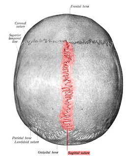
Suture in Human Head
Insect Sclerites
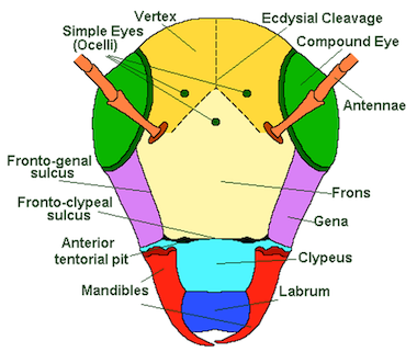
- Vertex: It is the top portion of the head behind the frons or the area between the two compound eyes.
- Frons: It is the facial part of the insect consisting of median ocellus.
- Clypeus: It is situated above the labrum and is divided into anterior ante-clypeus and posterior post-clypeus.
- Labrum: It is small sclerite that forms the upper lip of the mouth cavity. It is freely attached or suspended from the lower margin of the clypeus.
- Gena: It is the area extending from below the compound eyes to just above the mandibles.
- Occular sclerites: These are cuticular ring like structures present around each compound eye.
- Antennal sclerites: These form the basis for the antennae and present around the scape which are well developed in Plecoptera (stone flies).
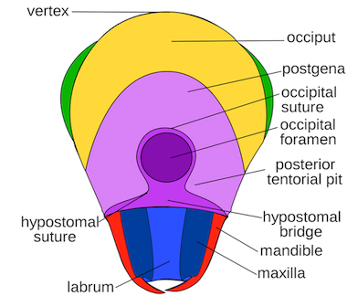
- Epicranium: It is the upper part of the head extending from vertex to occipital suture.
- Occiput: It is an inverted “U” shaped structure representing the area between the epicranium and post occiput.
- Post occiput: It is the extreme posterior part of the insect head that remains before the neck region.
All the above sclerites gets attached through cuticular ridges or sutures to provide the attachment for the muscles inside.
Head Sutures
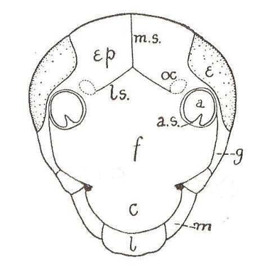
a. antennal socket; a.s. antennal sclerite; c. clypeus; e. compound eye; Ep. epicranial plate; f. frons; g. gena; l. labrum; ls. lateral arms of epicranial suture; m. mandible; m.s. median epicranial suture; oc. ocellus
- Sutures: Latin Word: means joint, helps in muscle attachment.
- Epicranial suture:
- It is an
inverted Y shapedsuture distributed above the facial region extending up to the epicranial part of the head. It consists of two arms called frontal suture occupying the frons and stem called as coronal suture. - This epicranial suture is also known as line of weakness or ecdysial suture because the exuvial membrane splits along this suture during the process of ecdysis (breakage of cuticle).
- It is an
- Clypeolabral suture: It is the suture present between clypeus and labrum. It remains in the lower margin of the clypeus from which the labrum hangs down.
- Clypeofrontal suture or epistomal suture: The suture present between clypeus and frons.
- Occipital suture:
- It is ‘U’ shaped or horseshoe shaped suture between epicranium and occiput.
- Occiput means behind the head.
- Post occipital suture:
- It is the
only real suturein insect head. Posterior end of the head is marked by the post occipital suture to which the sclerites are attached. - As this suture separates the head from the neck, hence named as real suture.
- It is the
- Genal suture: It is the sutures present on the lateral side of the head i.e. gena.
- Occular suture: It is circular suture present around each compound eye.
- Antennal suture: It is a marginal depressed ring around the antennal socket.
Head
- Posterior opening of the cranium through which aorta, foregut, central neve cord and neck muscles passes is known as occipital foramen.
- Endoskeleton of insect cuticle provides space for attachments of muscles of antenna & mouthparts, called as tentorium.
- The appendages like a pair of compound eyes, 0-3 ocelli, a pair of antenna & mouth parts are called as cephalic appendages.
Functions of Head
- Food ingestion
- Sensory perception
- Coordination of bodily activities
- Protection of the coordinating centres
Types of Head Orientations
- The orientation of head with respect to the rest of the body varies.
- According to the position or projection of mouth parts, the head of the insect can be classified as:
-
Hypognathous
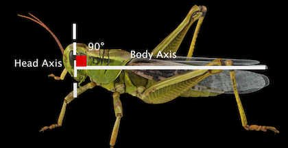
- Hypo – below: Gnathous – Jaw
- The head remain vertical and is at right angle to the long axis of the body and mouth parts are ventrally placed and projected downwards.
- This is also known as Orthopteroid type.
- This is the primitive condition.
- Eg: Grasshopper, Cockroach
-
Prognathous
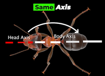
- Pro – infront: Gnathous – Jaw
- The head remains in the same axis to body and mouth parts are projected forward.
- This is common in insects that burrow and in predatory insects.
- This is also known as Coleopteroid type.
- Eg: Beetles
-
Opisthognathous
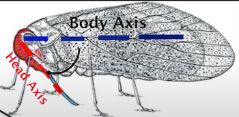
- Opistho – behind: Gnathous – Jaw
- It is same as prognathous, but mouthparts are directed back ward and held in between the fore legs.
- This is also known as Hemipteroid or Opisthorhynchous type.
- This condition found in sucking insects.
- Eg: Bugs
Antennae
- Single pair of antennae is present in most of insects.
- Antenna is consisting of three parts i.e., scape (basal part), pedicel and flagella.
- Antennae are a pair of sensory preoral appendages arising from the 2nd or antennal segment of the head possessing nerves coming from deutocerebrum of the brain.
- They are well developed in adults and poorly developed in immature stages.
- The base of socket is connected to the edge of the socket by an articulatory membrane. This permits free movement of antennae.
Structure of Antenna
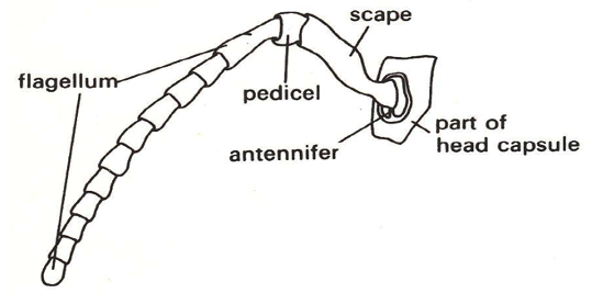
A typical Antenna of Pterygote Insect
- Antenna consists of 3 parts:
- Scape: It is the first segment of antenna. It articulates with the head capsule through antennifer which provides movement for the scape.
- Pedicel: It is the 2nd or middle segment of antenna that forms a joint between scape and flagellum. It consists of the special auditory organ known as
Jhonston’s organ. (As discovered by American Physician Christopher Johnston). - Flagellum: It is the last antennal segment which consists of many segments that varies in shape and size.
Modification of Antennae
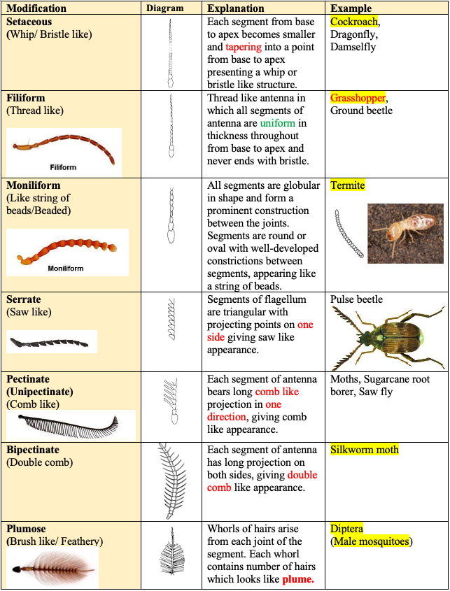
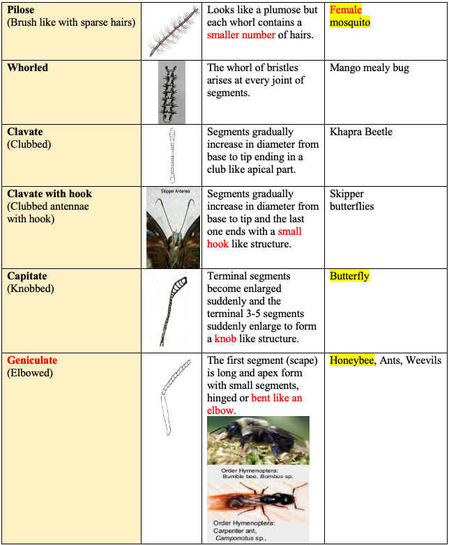
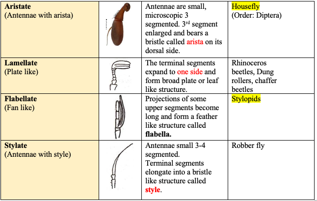
Functions of Antenna
- Mainly serve as the sense organ responding to touch, smell, odour, humidity change, variation in temperature, vibration, wind velocity and direction.
- Antenna is useful to detect chemicals including food and pheromones (chemicals secreted into air by opposite sex). The antennae of male silk moths are extremely sensitive to the female sex pheromone such that a male moth can find a female up to 4.5 km away. This remarkable sensitivity is due to both the morphological and biochemical design of these antennae.
- Antenna is useful to perceive the forward environment and detect danger.
- Jhonston’s organ on pedicel functions as an auditory organ responding to sound (helps in hearing) and helpful for measuring the speed of air currents. (Mainly found in male mosquito)
- In some insects, sound producing organ (mole crickets) are found in second segment of antenna.
- Helps the mandibles for holding prey and for mastication of food material.
- Helps in sexual dimorphism. (Distiction between male ⚦ and female ♀)
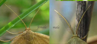
Sexual Diamorphism in Insects
- Useful for clasping the female during copulation.
- Aid in respiration by forming an air funnel in aquatic insects.
References
- Insecta - Introduction: K.N. Ragumoorithi, V. Balasurbramani & N. Natarajan
- A General Textbook of Entomology (9th edition, 1960) – A.D. Imms (Revised by Professor O.W. Richards and R.G. Davies). Butler & Tanner Ltd., Frome and London.
- The Insects- Structure and Function (4th Edition, 1998) – R.F. Chapman. Cambridge University Press
- Wikipedia
Chordoton organ found in Male Mosquito?
Insect Head
- Insect body is divided into three regions or tagmata namely head, thorax and abdomen.
- This grouping of body segments into regions is known as tagmosis.
- Insect head is a hard and highly sclerotized compact structure.
- Head consists of compound eyes, simple eyes (ocelli), mouthparts and a pair of antennae.
- It is the foremost part in insect body consisting of 7 segments that are fused to form a head capsule.
- The head segments can be divided into two regions i.e., procephalon and gnathocephalon (bear mouth parts).

Head Sclerites
- Head capsule is sclerotized and the head capsule excluding appendages formed by the fusion of several scleritis is known as cranium.
- It is hard and highly sclerotized compact structure.
- The head capsule is …
Become Successful With AgriDots
Learn the essential skills for getting a seat in the Exam with
🦄 You are a pro member!
Only use this page if purchasing a gift or enterprise account
Plan
- Unlimited access to PRO courses
- Quizzes with hand-picked meme prizes
- Invite to private Discord chat
- Free Sticker emailed
Lifetime
- All PRO-tier benefits
- Single payment, lifetime access
- 4,200 bonus xp points
- Next Level
T-shirt shipped worldwide

Yo! You just found a 20% discount using 👉 EASTEREGG

High-quality fitted cotton shirt produced by Next Level Apparel