🦋 Wing Structure
Analogy with other organisms, Wing Internals, Wing Venation, Margins and Angles, Wing Regions
Which of the following appendages found in Pterothorax region?
- Wings in insect gives it to a competitive advantage:
- To seek food, mate, shelter and oviposition sites
- To colonize in a new habitat and also to exchange habitat.
- To escape from enemies and unfavourable conditions.
- To migrate (i.e. for long distance travel e.g. Locusts)
- Insects were the first to evolve flight, approximately 350 million years ago. Even before dinosaurs. Pterosaurs were the next to evolve flight, approximately 228 million years ago.
Analogy with other organisms
- Insects are the only invertebrates possessing wings and capable of true flight.
- The wing of an insect and the wing of a bird are analogous organs. Both these organs are used for flying in the air, but they are very different in structure.
- An insect wing is an extension of the integument, whereas birdwing is formed of limb bones covered with flesh, skin, and feathers.

Insect Wings
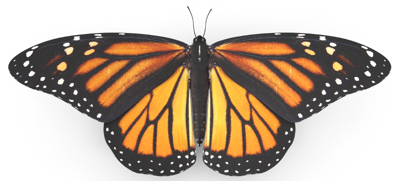
- Generally, insects has two pairs of wings are present in meso (second) and meta (third) thorax (pterothorax).
- Among the pterygotes, normally, two pairs of wings are present in insects, and they are borne on pterothoracic segments viz., mesothorax and metathorax.
- Front pair (First Pair) of wings are known as forewings and back pair of wings (Second Pair) are known as hind wings.
- Greek word pteron means wing.
- Insects originated from their primitive ancestors which were wingless.
- Based on the presence or absence of wings, class insects are divided into two subclasses.
- Apterygota
- Pterygota
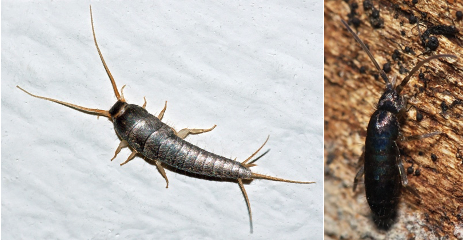
- The primitive apterygotes are primarily wingless. Eg: Silver fish and Spring tails.
- Ectoparasites like head louse, poultry louse and flea are secondarily wingless. That is, insect groups whose ancestors once had wings but that have lost them as a result of subsequent evolution. (Because they have to remain attached to their host so do not require wings)
- Wings are only present in adult stage.
- Wings are deciduous in ants and termites.
- There is only one pair of wings in the true flies. But in some insects second pair of wings (hind wings) are reduced.
Wing Internals
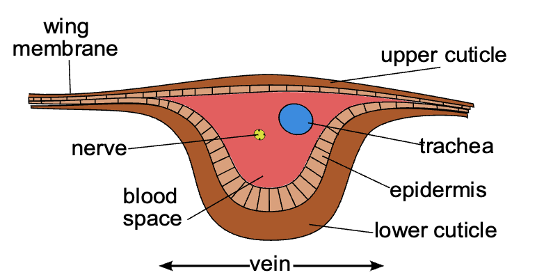
- Wing is a flattened double - layered expansion of body wall (exoskeleton) with a dorsal and ventral lamina having the same structure as the integument. Both dorsal and ventral laminae grow, meet and fuse except along certain lines. Thus, a series of channels is formed.
- These channels serve for the passage of tracheae, nerves and blood. Wing is nourished by blood circulating through veins. Later the walls of these channels become thickened to form veins or nerves.
- Wings are very thin broad leaf like structures strengthened by a number of hollow narrow tubular structures called veins.
Wing Venation
The arrangement (number and position) of veins on the wings is called wing venation which is extensively used in insect classification.
- The patterns resulting from the fusion and cross-connection of the wing veins are often diagnostic for different evolutionary lineages and can be used for identification to the family or even genus level in many orders of insects. Entomologists study the venation of wings and this is often used as a way of differentiating between otherwise similar species.
- Insect wings are subjected to considerable variations in shape, size, marking, and vein patterns reflecting their specific functional differences.
- Venation patterns are the most characteristic structures of wings because of aerodynamic importance associated with specific flight system and of specialized functional purposes, and there are marked differences between forewings and hindwings in a species.
Hypothetical Wing Venation
- These veins (and their branches) are named according to a system devised by John Comstock and George Needham — The Comstock–Needham system.
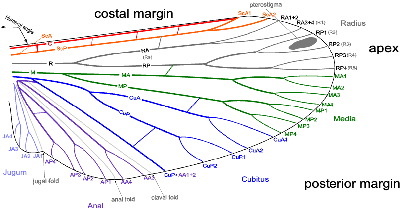
- Wing venation, which consists of two types of veins:
- Longitudinal Veins: Extend from base of the wing to the margin. They may be convex (crest, ∩) or concave (trough, U).
- Cross Veins: That interlink the longitudinal veins.
- Insect wing veins are composed of longitudinal veins and crossveins. The longitudinal veins are heavily sclerotized, providing conduits for nerves, the tracheae and circulating haemolymph, running alternately on the crests (convexes) or in the troughs (concaves) of wings.
- Their patterns are basically shared among extant winged insects and named after the Comstock and Needham system with some modifications based on comparative studies of extinct and extant insects.
- The principal six longitudinal veins arranged in order from the anterior margin are costa (C), sub costa (Sc), radius (R), median (M), cubitus (Cu) and anal veins (A).
- The veins of the wing appear to fall into an undulating pattern according to whether they have a tendency to fold up or down when the wing is relaxed.
- Costa (C): It forms the thickened anterior margin of the wing (costal) and is un-branched and is convex. Costa C, meaning rib.
- Sub costa (Sc): It runs immediately below the costa always in the bottom of a trough between C and R. It is forked distally. The two branches of Sc are Sc1 and Sc2 and is concave. Subcosta Sc, meaning below the rib.
- Radius vein (R): It is the next main vein, stout and connects at the base with second auxiliary sclerite, it divided in to two branches R1 and Rs (Radial sector). Radius R, in analogy with a bone in the forearm, the radius. R1 goes directly towards apical margin and is convex; Rs is concave and divided in to 4 branches, R2, R3, R4, R5.
- Media (M): It is one of the two veins articulating with some of the small median sclerites. Media M, meaning middle. It is divided in two branches: i) Media anterior (MA) which is convex and ii) Media posterior (MP) and is concave.
- Cubitus (Cu): Cubitus Cu, meaning elbow. It articulates with median auxiliary sclerite. Cubitus is divided into convex CU1 and concave CU2. CU1 is again divided into CU1a and CU1b.
- Anal veins (A): Anal veins A, in reference to its posterior location. These veins are convex. They are individual un-branched, 1-3 in number.
👉🏻 There is another small vein known as Jugal veins. 1 or 2 jugal veins (unbranched) are present in the jugal lobe of the forewing.
Cross Veins
Small veins often found interconnecting the longitudinal veins are called cross veins.
- Large numbers of cross-veins are present in some insects, and they may form a reticulum as in the wings of Odonata (dragonflies and damselflies).
- In early insects the veins running down the wing (longitudinal veins) were connected by a series of cross veins. Most insect groups have lost, or dramatically reduced the number of, these cross veins. However, some insects such as dragonflies and damselflies have wings that contain many cross veins.
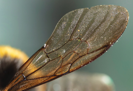
- Cross veins reduced in advanced insect orders Example Bumblebee’s wing.
- Ordinarily, however, there is a definite number of cross-veins having specific locations. The more constant cross-veins are the humeral cross-vein (h) between costa and subcosta, the radial cross-vein (r) between R and the first fork of Rs, the sectorial cross-vein (s) between the two forks of R8, the median cross-vein (m–m) between M2 and M3, and the mediocubital cross-vein (m-cu) between media and cubitus.
- Humeral cross vein (h): between costa and subcosta
- Radial cross vein (r): between subcostal and radius R1
- Sectorial cross veins (s): between subbranches of radial sector
- Radio medial cross vein (r-m): between radius and media
- Median cross veins (m-m): between branches of media
- Medio-cubital veins (m-cu): between media and cubitus
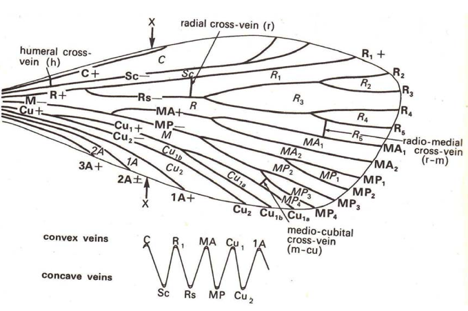
Hypothetical Wing Venation
Margins and Angles
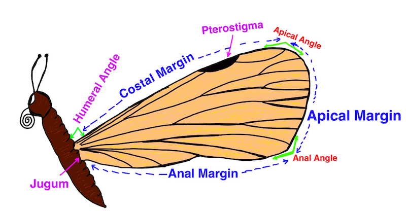
- A typical insect wing is triangular with three margins and three angles.
- The wing is triangular in shape and has therefore three sides/margins and three angles.
- Three margins are:
- Costal or anterior
- Apical or outer and
- Anal or inner
- The anterior margin strengthened by the costa is called coastal margin and the lateral margin is called apical margin and the posterior margin is called anal margin.
- Three angles are:
- Apical angle or outer angle: between costal margin and apical margin
- Anal angle: between apical and anal margin
- Humeral angle: The angle by which the wing is attached to the thorax or angle between body wall and costal margin is called humeral angle.
Wing Regions
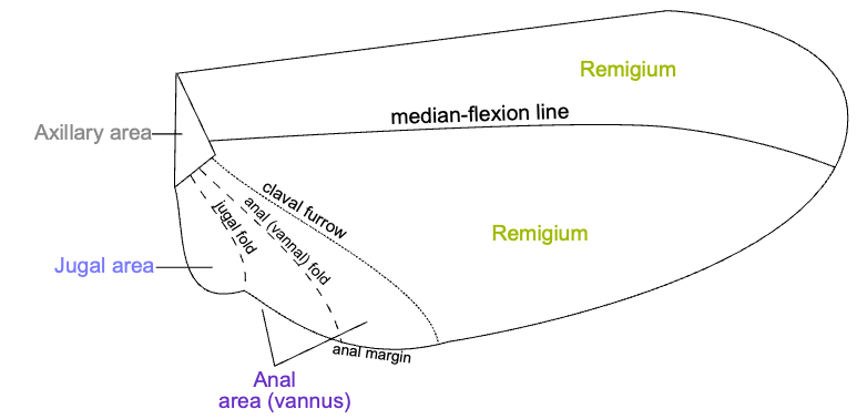
- The surface area of typical insect wing is divided in to two portions i.e., Remegium and Vannal Area.
- The anterior (upper) part of the wing towards coastal margin where a greater number of longitudinal veins are present is called remigium. Most veins and crossveins occur in the anterior area of the remigium, which is responsible for most of the flight, powered by the thoracic muscles.
- The flexible posterior part of the wing where veins are sparsely distributed is known as vannal area, which is called as clavus in forewings and vanus in hindwings.
- Jugum is the inner most portion of the wing that is cutoff from the main wing by jugal fold. Means the two regions are separated by jugal fold.
- The proximal part of vannus is called jugum, when well-developed is separated by a jugal fold.
- The insect wings may sometimes possess some pigmented spot near coastal margin known as pterostigma or stigma as in Odonata (dragon flies and damsel flies).
- The area containing wing articulation sclerites is called axilla.
- Each wing is attached to the body by a membranous basal area, but the articular membrane contains a number of small articular sclerites, collectively known as the pteralia.
- Wings are moved by thoracic flight muscles attached to their bases.
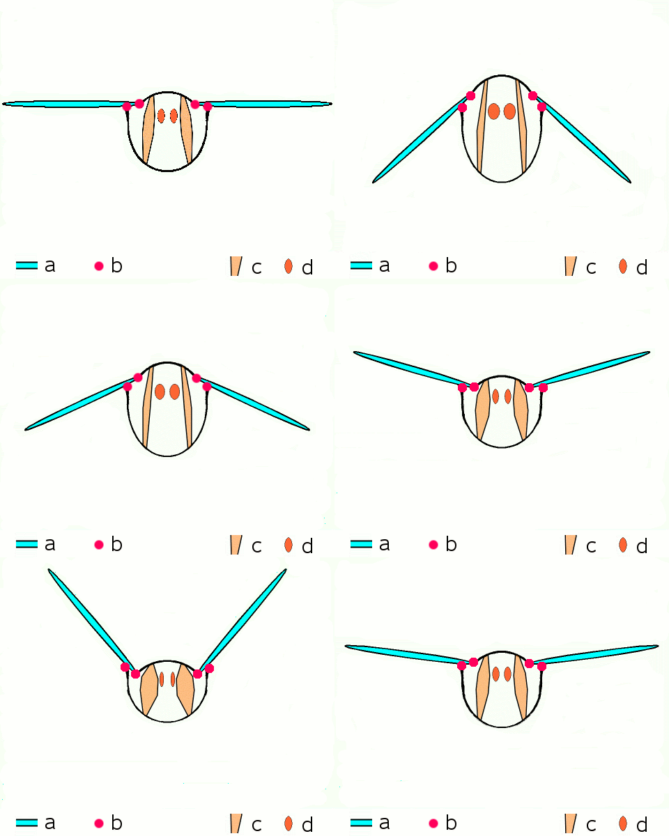
Motion of an insect wing: a. wings b. primary and secondary flight joints c. dorsoventral flight muscles d. longitudinal muscles
References
- Insecta - Introduction: K.N. Ragumoorithi, V. Balasurbramani & N. Natarajan
- A General Textbook of Entomology (9th edition, 1960) – A.D. Imms (Revised by Professor O.W. Richards and R.G. Davies). Butler & Tanner Ltd., Frome and London.
- The Insects- Structure and Function (4th Edition, 1998) – R.F. Chapman. Cambridge University Press
- Wikipedia
Which of the following appendages found in Pterothorax region?
- Wings in insect gives it to a competitive advantage:
- To seek food, mate, shelter and oviposition sites
- To colonize in a new habitat and also to exchange habitat.
- To escape from enemies and unfavourable conditions.
- To migrate (i.e. for long distance travel e.g. Locusts)
- Insects were the first to evolve flight, approximately 350 million years ago. Even before dinosaurs. Pterosaurs were the next to evolve flight, approximately 228 million years ago.
Analogy with other organisms
- Insects are the only invertebrates possessing wings and capable of true flight.
- The wing of an insect and the wing of a bird are analogous organs. Both these organs are used for flying in the air, but they are very different in structure.
- An …
Become Successful With AgriDots
Learn the essential skills for getting a seat in the Exam with
🦄 You are a pro member!
Only use this page if purchasing a gift or enterprise account
Plan
- Unlimited access to PRO courses
- Quizzes with hand-picked meme prizes
- Invite to private Discord chat
- Free Sticker emailed
Lifetime
- All PRO-tier benefits
- Single payment, lifetime access
- 4,200 bonus xp points
- Next Level
T-shirt shipped worldwide

Yo! You just found a 20% discount using 👉 EASTEREGG

High-quality fitted cotton shirt produced by Next Level Apparel