🫁 Anatomy
Learn about inner body structure of plant parasitic nematodes and different variations in stoma & oesophagus
Which of the following is correct regarding nematode?
👉🏻 The inner body tube of nematode comprises of the digestive system which is divisible into three distinct zones:
- Stomodeum/Foregut: which constitute the stoma, oesophagus and cardia.
- Mesenteron/Midgut: which constitute the intestine.
- Proctodaeum/Hindgut: which is the posterior–most region comprising rectum and anal opening.
Stomodeum (Fore Gut)
- It is the anterior part of alimentary canal.
- Stomodeum is primarily responsible for feeding (intake of food and partial digestion of food).
- Parts of stomodeum are:
- Cephalic region
- Stoma
- Oesophagus
- Oesophageal glands
- Cardia
Cephalic Region
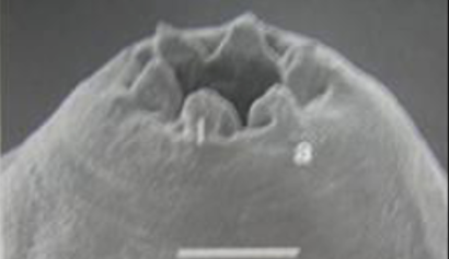
- It constitutes oral opening, lips, cephalic framework and basal ring.
- Oral Opening: It is opening of mouth surrounded by the six lips.
- Lips: There are six lips which are
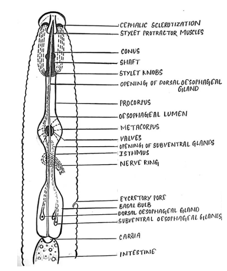
hexaradiallyarranged with oral opening at the centre. - Cephalic Framework: Below each lip, there is an inverted basket like sclerotised radial muscles which are hexaradially arranged called cephalic framework. The cephalic framework gives support to the lips during feeding.
- Basal Ring: Base of the cephalic framework is called basal ring on which one end of the protractor muscles are attached and the other end to the base of the stylet knobs. These protractor muscles help in forward and backward movement of stylet during insertion into the plant cell for feeding.
Stoma
- Stoma is the portion of the inner body tube lying between the oral opening and oesophagus.
- The stomatal opening is small and slit like and is surrounded by six lips.

- Plant parasitic nematodes are armed with a protrusible stylet which is usually hallow and functions like a hypodermic needle.
- In, Secernentea (Order of Plant Parasitic Nematodes), the stylet is thought to be derived from fusion of the stomatal lining and therefore called as
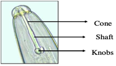
stomatostylet. Stomatostylet developed from fusion of walls of buccal cavity. - It used for piercing into plant cells. The stamatostylet consists of a anterior cone, a cylindrical shaft and three rounded basal knobs.

- In Adenophrea, the stylet is thought to be derived from a tooth and, therefore, it is called as odontostylet.
- Stylet functions to pierce the cell wall of the root.
- The nematode secrets a hallow tube out of its stoma that connect it with the plant.
- This feeding tube serves as the interface between the nematode and the plant.
Oesophagus
- Oesophagus is found in between the stoma and intestine.
- Mainly responsible for transport of food by pumping.
- It prevents the regurgitation.
- Depending upon the diversity in feeding habits among different groups of nematodes, oesophagus is of three types.
- One part oesophagus
- Two-part oesophagus
Three-part oesophagus: Found in Plant Parasitic Nematodes. Contains three parts - Corpus, Isthmus and Basal bulb.
- Oesophageal Glands: Digestive juices secreted from glands are injected into the host plant cell by means of the stylet. Helps in digestion.
Cardia
- Cardia is the posterior part of stomodeum.
- It is otherwise called as
Oesophago-Intestinal junction. - Cardia comprises of a triradiate valve which controls the passage of food from oesophagus to intestine preventing regurgitation (back flow to oesophagus).

Mesenteron (Mid Gut)
- It is the middle part of the digestive system called the Intestine.
- It is a hollow straight tube lined with single layer of endodermal epithelial cells.
- Initima (cuticular lining) is absent and fine finger like projections are known as
microvillipresent around the lumen. - The microvilli are finger like projection of the plasma membrane projecting in to the intestinal regions.
- They increase the surface area of the intestine and are both secretary and absorbtive in function.

Proctodaeum (Hindgut)
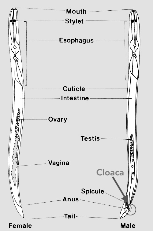
- Proctodeum comprises rectum and anus.
- The intestinal tube is connected with a narrow small tube at the posterior end, through a valve known as rectum.
- It regulates the flow of undigested food material which is to be passed outside the nematode body through a ventrally located aperture known as anus.
- In
male nematode, the rectum joins with the hind part of the testis forming a common opening known ascloaca. - In female, there is a separate opening.
- Oesophageal and rectal glands are present in nematodes. The oesophageal gland enter the stomodeum and rectal gland enter proctodeum.

References
- Walia, R. K and Bajaj, H. K (2014). Textbook of Introductory Plant Nematology. Directorate of Knowledge Management in Agriculture, ICAR, New Delhi.
- Ravichandra, N. G. (2019). Plant Nematology. I. K. International Publishing House Pvt. Ltd., New Delhi.
- Dasgupta, M. K. (1998). Phytonematology. Pilgrims Publishing
- Fotedar, D.N. & Handoo, Z.A. (1978) A revised scheme of classification to order Tylenchida Thorne, 1949 (Nematoda). Journal of Science, University of Kashmir (1975), 3, 55–82.
- Qing, X., Bert, W. Family Tylenchidae (Nematoda): an overview and perspectives. Org Divers Evol 19, 391–408 (2019).
- https://doi.org/10.1007/s13127-019-00404-4
- https://nematode.unl.edu/dolichod.htm
- http://www.nematologia.com.br/files/tematicos/6.pdf
Which of the following is correct regarding nematode?
👉🏻 The inner body tube of nematode comprises of the digestive system which is divisible into three distinct zones:
- Stomodeum/Foregut: which constitute the stoma, oesophagus and cardia.
- Mesenteron/Midgut: which constitute the intestine.
- Proctodaeum/Hindgut: which is the posterior–most region comprising rectum and anal opening.
Stomodeum (Fore Gut)
- It is the anterior part of alimentary canal.
- Stomodeum is primarily responsible for feeding (intake of food and partial digestion of food).
- Parts of stomodeum are:
- Cephalic region
- Stoma
- Oesophagus
- Oesophageal glands
- Cardia
Cephalic Region
- It constitutes oral opening, lips, cephalic framework and basal ring.
- Oral Opening: It is opening of mouth surrounded by the six lips.
- Lips: There …
Become Successful With AgriDots
Learn the essential skills for getting a seat in the Exam with
🦄 You are a pro member!
Only use this page if purchasing a gift or enterprise account
Plan
Rs
- Unlimited access to PRO courses
- Quizzes with hand-picked meme prizes
- Invite to private Discord chat
- Free Sticker emailed
Lifetime
Rs
1,499
once
- All PRO-tier benefits
- Single payment, lifetime access
- 4,200 bonus xp points
- Next Level
T-shirt shipped worldwide

Yo! You just found a 20% discount using 👉 EASTEREGG

High-quality fitted cotton shirt produced by Next Level Apparel