🧖🏽♀️ Morphology -- Outer Body Structure
Cuticle, hypodermis, somatic muscles, and sensory structures (amphid, phasmid, deirid) of plant parasitic nematodes
In the previous lesson, we covered the general body organisation of nematodes -- the tube-within-a-tube plan, body shape, and symmetry. Now we zoom into the outer tube itself: the three-layered body wall and its sensory structures.
How does a root-knot nematode find a tomato root in a vast expanse of soil? How does it squeeze through tiny soil pores to reach its target? The answers lie in the nematode's outer body wall -- a sophisticated structure equipped with sensory organs for host detection, a flexible yet protective cuticle, and muscles for movement.
Think of nematode morphology like a submarine: The cuticle is the hull (tough, protective, flexible). The amphids at the front are the sonar system (detecting chemical signals from roots). The phasmids at the rear are rear sensors. The hypodermis is the inner lining that maintains the hull. And the stylet (in plant parasites) is the drill bit that penetrates into roots.
Pro Content Locked
Upgrade to Pro to access this lesson and all other premium content.
₹99 charged monthly · Cancel anytime
- All Agriculture & Banking Courses
- AI Lesson Questions (100/day)
- AI Doubt Solver (50/day)
- Glows & Grows Feedback (30/day)
- AI Section Quiz (20/day)
- 22-Language Translation (100/day)
- Recall Questions (20/day)
- AI Quiz (15/day)
- AI Quiz Paper Analysis (100/day)
- AI Step-by-Step Explanations (100/day)
- Spaced Repetition Recall (FSRS)
- AI Tutor
- Immersive Text Questions
- Audio Lessons — Hindi & English
- Mock Tests & Previous Year Papers
- Summary & Mind Maps
- XP, Levels, Leaderboard & Badges
- Generate New Classrooms
- Voice AI Teacher (AgriDots Live)
- AI Revision Assistant
- Knowledge Gap Analysis
- Interactive Revision (LangGraph)
🔒 Secure via Razorpay · Cancel anytime · No hidden fees
In the previous lesson, we covered the general body organisation of nematodes -- the tube-within-a-tube plan, body shape, and symmetry. Now we zoom into the outer tube itself: the three-layered body wall and its sensory structures.
How does a root-knot nematode find a tomato root in a vast expanse of soil? How does it squeeze through tiny soil pores to reach its target? The answers lie in the nematode's outer body wall -- a sophisticated structure equipped with sensory organs for host detection, a flexible yet protective cuticle, and muscles for movement.
Think of nematode morphology like a submarine: The cuticle is the hull (tough, protective, flexible). The amphids at the front are the sonar system (detecting chemical signals from roots). The phasmids at the rear are rear sensors. The hypodermis is the inner lining that maintains the hull. And the stylet (in plant parasites) is the drill bit that penetrates into roots.
This lesson covers:
- Cuticle -- the non-living outer covering, its layers, markings, and functions
- Sensory structures -- amphid, phasmid, deirid, and caudal alae
- Hypodermis -- the living layer that secretes and maintains the cuticle
- Somatic muscles -- the locomotion machinery
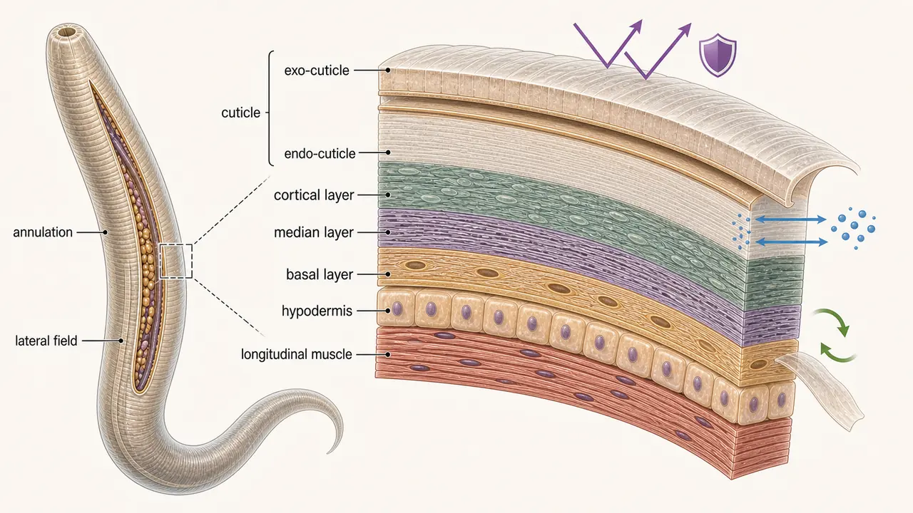
The Three Layers of the Body Wall
The outer body tube (body wall) consists of three layers, from outside to inside. Each layer performs distinct functions critical for nematode survival in soil.
The outer body tube (body wall) consists of three layers, from outside to inside:
| Layer | Nature | Primary Function |
|---|---|---|
| Cuticle (outermost) | Non-living, non-cellular | Protection, shape, sensory perception |
| Hypodermis (middle) | Single living cellular layer | Cuticle secretion, osmotic regulation |
| Somatic longitudinal muscles (innermost) | Muscle tissue rich in glycogen | Movement |
Cuticle
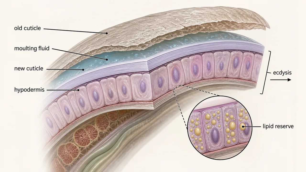
The cuticle is a non-living, non-cellular, triple-layered covering secreted by the underlying hypodermis. Because it is non-living, it cannot grow or repair itself -- it must be periodically shed and replaced through moulting (ecdysis).
Structure
The cuticle covers the entire body and lines the oesophagus, vulva, anus, cloaca, excretory pore, and sensory organs. Two important cuticular derivatives are:
- Feeding stylet -- used to pierce plant cells
- Copulatory spicules -- used by males during mating
The cuticle consists of three primary zones:
| Zone | Position |
|---|---|
| Cortical layer | Outermost |
| Median layer | Middle |
| Basal layer | Innermost |
The external lining is called exo-cuticle and the internal lining is called endo-cuticle.
Cuticular Markings
Surface markings on the cuticle serve as taxonomic fingerprints for species identification:
| Type | Description |
|---|---|
| Punctuation | Dot-like markings |
| Transverse striations | Ring-like markings around the body |
| Longitudinal markings | Lines running along the body length |
Functions of the Cuticle
- Exoskeleton: Together with the hypodermis, forms the exoskeleton that provides body shape (works with pseudocoelom as a hydrostatic skeleton).
- Protection: Shields the nematode from harsh soil environments.
- Movement: Provides the structural basis for sinusoidal locomotion through soil and plant tissue.
- Gas exchange: Since nematodes lack a respiratory system, gas exchange occurs by diffusion across the cuticle.
- Permeability: Acts as a semipermeable membrane -- allows water and non-electrolytes in, blocks undesirable elements.
- Sensory response: Contains sensory structures that detect external stimuli.
- Growth via moulting: Nematodes undergo four moults from J1 to adult. In each moult, both exo- and endo-cuticle are replaced by new cuticle secreted from the hypodermis -- a process called ecdysis.
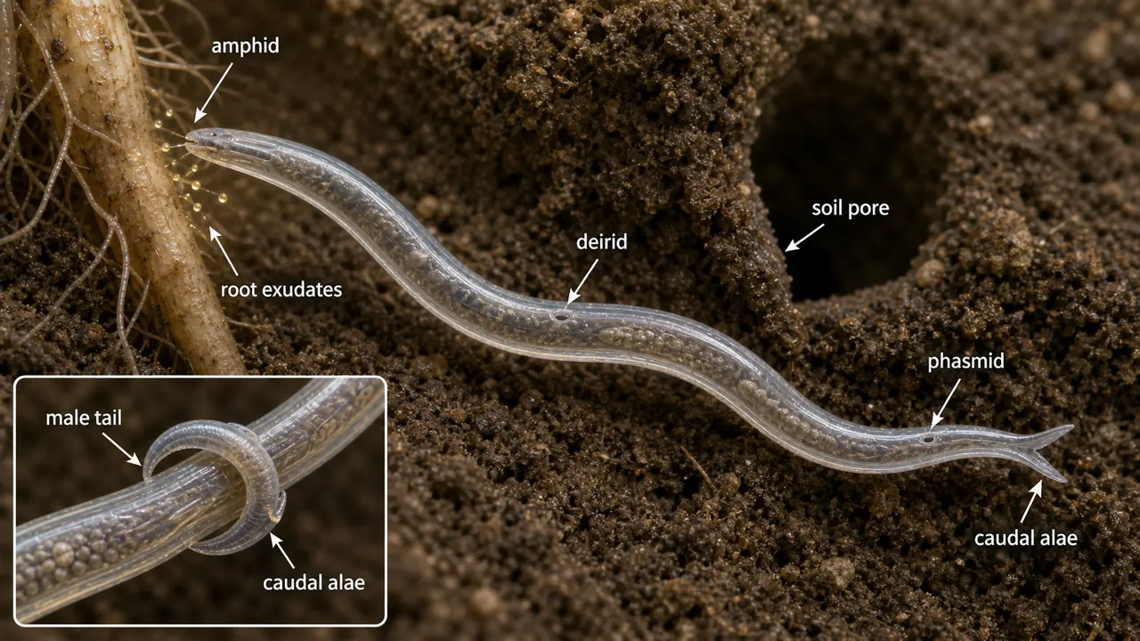
Sensory Structures of the Cuticle
Three pairs of sensory structures are associated with the cuticle surface. These are key diagnostic features and frequently tested in exams.
IMPORTANT
Amphid = chemoreceptor (host detection), Phasmid = chemoreceptor (aggregation/scent trail), Deirid = mechanoreceptor (pore size detection).
Amphid
- A pair of lateral chemosensory structures on the surface of the cuticle at the lateral lips, positioned opposite each other.
- Function: Chemoreceptor that detects chemical changes in the environment, helping the nematode recognise a favourable host plant by detecting root exudates. Think of amphids as the nematode's sense of smell.
Phasmid
- A pair of lateral chemosensory structures appearing as pore-like openings in the mid-tail region, opposite each other.
- Function: Chemoreceptor that acts as a scent trail, attracting members of the same species to assemble together at feeding sites. This chemical aggregation explains why plant parasitic nematodes are often found in clusters around host roots.
NOTE
The presence or absence of phasmids is a fundamental taxonomic character: Class Secernentea (Phasmida) has phasmids; Class Adenophorea (Aphasmida) lacks them.
Deirid
- A pair of lateral outgrowths at the centre of the lateral field, just below the nerve ring.
- Function: Mechanoreceptor that determines the size of soil pore spaces before the nematode attempts to pass through. Think of deirids as the nematode's sense of touch, helping it assess whether a gap is large enough for its body.
Caudal Alae (Bursa)
- A pair of lateral, wing-like cuticular expansions around the tail of the male nematode.
- Commonly called the Bursa.
- Function: Helps the male grasp and clasp the female during mating for proper alignment of reproductive openings.
Hypodermis
The hypodermis is a single, living cellular layer lying beneath the non-living cuticle. It is metabolically the most active layer of the body wall.
| Function | Details |
|---|---|
| Cuticle secretion | Produces and maintains the cuticle throughout life |
| Moulting | Induces ecdysis by secreting leucine amino peptidase enzymes that break down the old cuticle |
| Osmotic and ionic regulation | Maintains water and ion balance for survival in varying soil moisture |
| Energy reservoir | Stores lipid reserves used during starvation or migration between hosts |
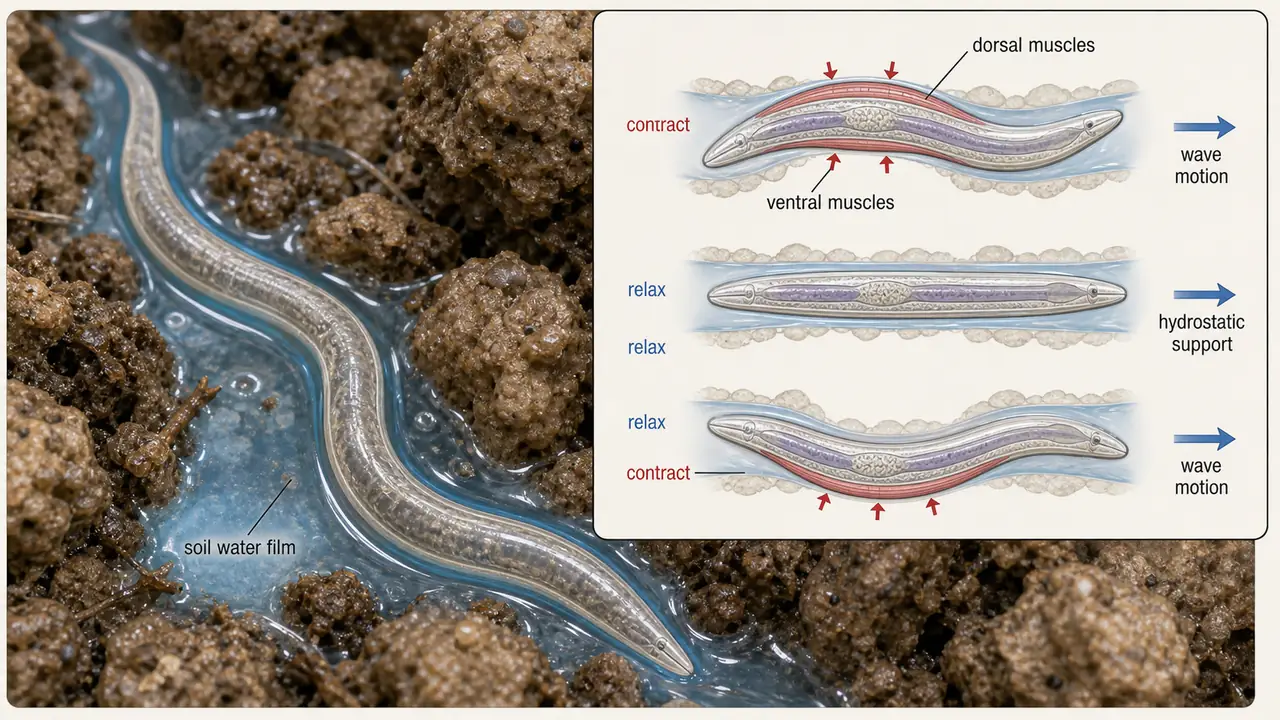
Somatic Longitudinal Muscles
The innermost lining of the body wall consists of somatic longitudinal muscles arranged in rows at the base of the hypodermis.
| Feature | Details |
|---|---|
| Arrangement | Run along the length of the body |
| Energy source | Rich in glycogen (stored sugar for quick energy during starvation) |
| Movement mechanism | Alternate contraction of dorsal and ventral muscle groups produces the characteristic sinusoidal wave motion |
Summary Table
| Structure | Nature | Key Function | Exam Tip |
|---|---|---|---|
| Cuticle | Non-living, non-cellular | Protection, exoskeleton, gas exchange | Three zones: cortical, median, basal; shed 4 times (ecdysis) |
| Amphid | Cuticular sensory organ | Chemoreceptor -- host plant detection | Located on lateral lips |
| Phasmid | Cuticular sensory organ | Chemoreceptor -- aggregation scent trail | Present in Secernentea, absent in Adenophorea |
| Deirid | Cuticular sensory organ | Mechanoreceptor -- pore size detection | Below nerve ring on lateral field |
| Caudal alae (Bursa) | Cuticular expansion | Male grasps female during mating | Male-only structure |
| Hypodermis | Living, single-cell layer | Cuticle secretion, moulting, energy storage | Secretes leucine amino peptidase for moulting |
| Somatic muscles | Longitudinal muscle rows | Sinusoidal movement | Rich in glycogen |
TIP
Exam mnemonic -- "APD" for sensory structures: Amphid = host-finding chemoreceptor (like a nose), Phasmid = aggregation chemoreceptor (like a pheromone sensor), Deirid = mechanoreceptor (like a touch sensor).
References
- Walia, R. K and Bajaj, H. K (2014). Textbook of Introductory Plant Nematology. Directorate of Knowledge Management in Agriculture, ICAR, New Delhi.
- Ravichandra, N. G. (2019). Plant Nematology. I. K. International Publishing House Pvt. Ltd., New Delhi.
- Dasgupta, M. K. (1998). Phytonematology. Pilgrims Publishing
- Fotedar, D.N. & Handoo, Z.A. (1978) A revised scheme of classification to order Tylenchida Thorne, 1949 (Nematoda). Journal of Science, University of Kashmir (1975), 3, 55-82.
- Qing, X., Bert, W. Family Tylenchidae (Nematoda): an overview and perspectives. Org Divers Evol 19, 391-408 (2019).
- https://doi.org/10.1007/s13127-019-00404-4
- https://nematode.unl.edu/dolichod.htm
- http://www.nematologia.com.br/files/tematicos/6.pdf
Summary Cheat Sheet
| Concept / Topic | Key Details |
|---|---|
| Cuticle (outermost) | Non-living, non-cellular — Protection, shape, sensory perception |
| Hypodermis (middle) | Single living cellular layer — Cuticle secretion, osmotic regulation |
| Somatic longitudinal muscles (innermost) | Muscle tissue rich in glycogen — Movement |
| Cortical layer | Outermost |
| Median layer | Middle |
| Basal layer | Innermost |
| Punctuation | Dot-like markings |
| Transverse striations | Ring-like markings around the body |
| Longitudinal markings | Lines running along the body length |
| Cuticle secretion | Produces and maintains the cuticle throughout life |
| Moulting | Induces ecdysis by secreting leucine amino peptidase enzymes that break down the old cuticle |
| Osmotic and ionic regulation | Maintains water and ion balance for survival in varying soil moisture |
| Energy reservoir | Stores lipid reserves used during starvation or migration between hosts |
| Arrangement | Run along the length of the body |
| Energy source | Rich in glycogen (stored sugar for quick energy during starvation) |
| Movement mechanism | Alternate contraction of dorsal and ventral muscle groups produces the characteristic sinusoidal wave motion |
| Cuticle | Non-living, non-cellular — Protection, exoskeleton, gas exchange — Three zones: cortical, median, basal; shed 4 times (ecdysis) |
| Amphid | Cuticular sensory organ — Chemoreceptor -- host plant detection — Located on lateral lips |
| Phasmid | Cuticular sensory organ — Chemoreceptor -- aggregation scent trail — Present in Secernentea, absent in Adenophorea |
| Deirid | Cuticular sensory organ — Mechanoreceptor -- pore size detection — Below nerve ring on lateral field |
| Caudal alae (Bursa) | Cuticular expansion — Male grasps female during mating — Male-only structure |
| Hypodermis | Living, single-cell layer — Cuticle secretion, moulting, energy storage — Secretes leucine amino peptidase for moulting |
| Somatic muscles | Longitudinal muscle rows — Sinusoidal movement — Rich in glycogen |
TIP
Next: Lesson 05 covers the inner tube -- the digestive system (anatomy) -- stoma, stylet types, three-part oesophagus, and the cloaca vs anus distinction.