🫁 Anatomy -- Digestive System
Inner body structure of nematodes -- stoma, stylet types, three-part oesophagus, intestine, and the male cloaca vs female anus distinction
In the previous lesson, we examined the outer body wall -- cuticle, hypodermis, muscles, and sensory structures. Now we move inward to the inner tube: the digestive system that enables nematodes to feed on plant cells.
When a root-knot nematode juvenile pierces a tomato root cell, it injects digestive enzymes through a hollow needle-like stylet and sucks back the pre-digested cell contents -- all through a structure barely visible under a microscope. This remarkable feeding apparatus is part of the nematode's digestive system, which runs as a complete alimentary canal from mouth to anus.
Think of the nematode digestive system like a syringe feeding system: The stylet is the needle — it pierces the plant cell wall. The oesophageal bulb is the pump — it sucks cell contents upward. The intestine is the absorption chamber. And the whole system is powered by muscular pumping, not gravity. This is why nematodes can feed from any angle underground.
Pro Content Locked
Upgrade to Pro to access this lesson and all other premium content.
₹99 charged monthly · Cancel anytime
- All Agriculture & Banking Courses
- AI Lesson Questions (100/day)
- AI Doubt Solver (50/day)
- Glows & Grows Feedback (30/day)
- AI Section Quiz (20/day)
- 22-Language Translation (100/day)
- Recall Questions (20/day)
- AI Quiz (15/day)
- AI Quiz Paper Analysis (100/day)
- AI Step-by-Step Explanations (100/day)
- Spaced Repetition Recall (FSRS)
- AI Tutor
- Immersive Text Questions
- Audio Lessons — Hindi & English
- Mock Tests & Previous Year Papers
- Summary & Mind Maps
- XP, Levels, Leaderboard & Badges
- Generate New Classrooms
- Voice AI Teacher (AgriDots Live)
- AI Revision Assistant
- Knowledge Gap Analysis
- Interactive Revision (LangGraph)
🔒 Secure via Razorpay · Cancel anytime · No hidden fees
In the previous lesson, we examined the outer body wall -- cuticle, hypodermis, muscles, and sensory structures. Now we move inward to the inner tube: the digestive system that enables nematodes to feed on plant cells.
When a root-knot nematode juvenile pierces a tomato root cell, it injects digestive enzymes through a hollow needle-like stylet and sucks back the pre-digested cell contents -- all through a structure barely visible under a microscope. This remarkable feeding apparatus is part of the nematode's digestive system, which runs as a complete alimentary canal from mouth to anus.
Think of the nematode digestive system like a syringe feeding system: The stylet is the needle — it pierces the plant cell wall. The oesophageal bulb is the pump — it sucks cell contents upward. The intestine is the absorption chamber. And the whole system is powered by muscular pumping, not gravity. This is why nematodes can feed from any angle underground.
Why anatomy matters for pest management: The stylet type (stomatostylet vs odontostylet) determines whether a nematode is a plant parasite or a predator/fungivore. Knowing this helps identify whether a nematode found in soil is actually a pest or a beneficial organism.
This lesson covers:
- Stomodeum (foregut) -- cephalic region, stoma, stylet types, three-part oesophagus, cardia
- Mesenteron (midgut) -- intestine with microvilli
- Proctodaeum (hindgut) -- rectum, anus vs cloaca distinction
Overview -- Three Regions of the Digestive System
The inner body tube is divisible into three distinct zones:
| Region | Alternative Name | Components | Function |
|---|---|---|---|
| Stomodeum | Foregut | Stoma, oesophagus, cardia | Food ingestion and initial processing |
| Mesenteron | Midgut | Intestine | Digestion and nutrient absorption |
| Proctodaeum | Hindgut | Rectum, anus | Waste expulsion |
Stomodeum (Foregut)
The anterior part of the alimentary canal, responsible for feeding. It contains the most complex structures involved in host-parasite interaction.
Cephalic Region
The feeding end of the nematode consists of four structures working together:
| Structure | Description |
|---|---|
| Oral opening | Mouth, surrounded by six lips |
| Lips | Six lips arranged in hexaradial symmetry around the oral opening |
| Cephalic framework | Inverted basket-like sclerotised radial muscles below each lip, giving rigidity during feeding |
| Basal ring | Base of the cephalic framework; one end of the protractor muscles attaches here, the other end to the stylet knobs, enabling forward-backward stylet thrust |
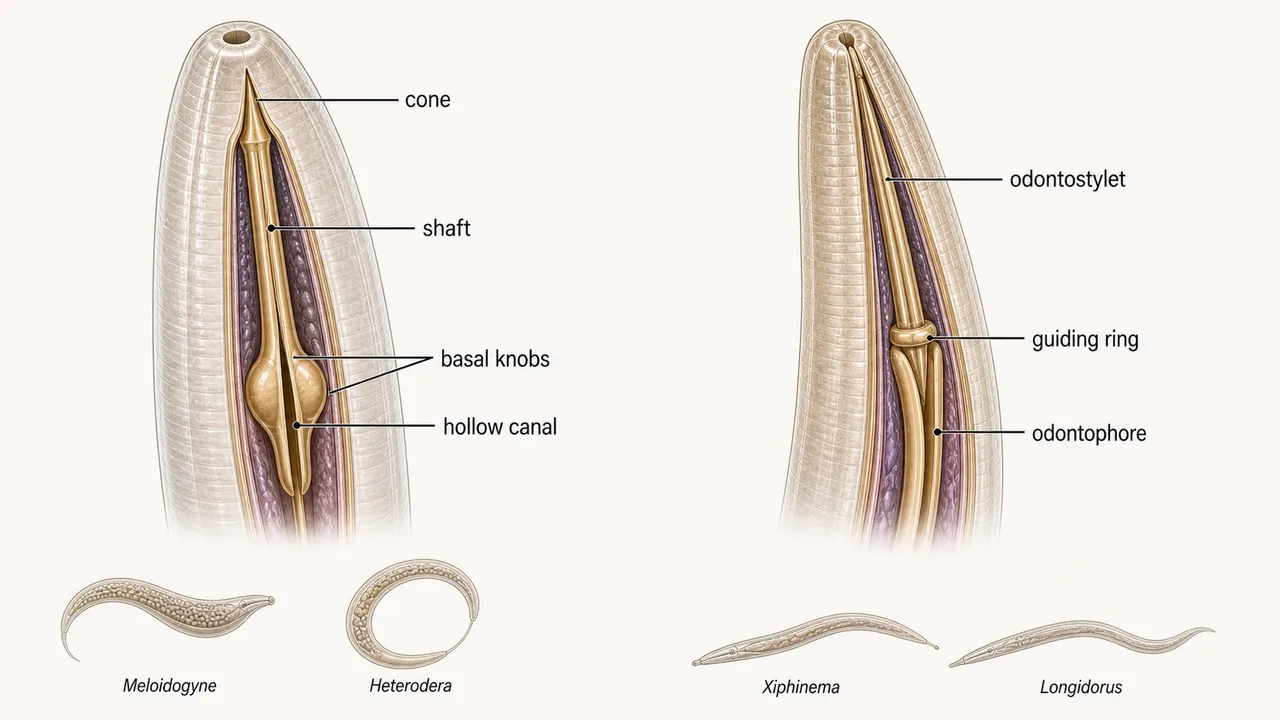
Stoma and Stylet
The stoma is the portion between the oral opening and oesophagus. In plant parasitic nematodes, it contains a protrusible stylet -- the most important diagnostic feature confirming a nematode is a plant parasite rather than a free-living species.
The stylet functions like a hypodermic needle: it pierces cell walls, injects digestive enzymes, and sucks back pre-digested cell contents.
Two Types of Stylet
| Feature | Stomatostylet | Odontostylet |
|---|---|---|
| Found in | Class Secernentea (Orders Tylenchida, Aphelenchida) | Class Adenophorea (Order Dorylaimida) |
| Origin | Derived from fusion of stomatal (buccal cavity) walls | Derived from a tooth |
| Parts | Anterior cone + cylindrical shaft + three rounded basal knobs | Modified tooth-like structure |
| Examples | Meloidogyne, Heterodera, Pratylenchus | Xiphinema, Longidorus (virus vectors) |
The nematode secretes a hollow feeding tube out of its stoma that connects it to the plant cell. Through this tube, enzymes flow in and pre-digested cell contents are sucked back -- a two-way traffic central to how nematodes derive nutrition.
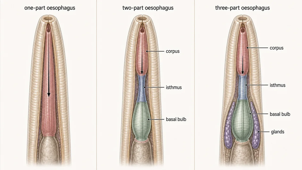
Oesophagus
Located between the stoma and intestine, the oesophagus acts as a muscular pump that generates suction to draw food through the narrow stylet. It also prevents regurgitation of food.
Three Types of Oesophagus
| Type | Found In |
|---|---|
| One-part oesophagus | Free-living nematodes |
| Two-part oesophagus | Some nematode groups |
| Three-part oesophagus | Plant parasitic nematodes (key diagnostic feature) |
The three-part oesophagus of plant parasitic nematodes consists of:
| Part | Description |
|---|---|
| Corpus | Anterior muscular portion |
| Isthmus | Narrow connecting tube |
| Basal bulb (posterior bulb) | Contains powerful pumping muscles and valve mechanisms |
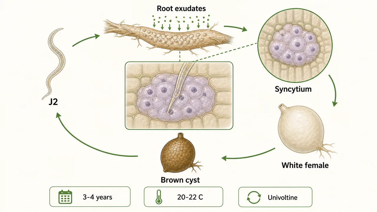
Oesophageal glands secrete digestive juices that are injected into the host cell via the stylet. These secretions break down cell walls and modify host cell metabolism -- this is how nematodes manipulate plant cells to form feeding sites like giant cells and syncytia.
Cardia (Oesophago-Intestinal Junction)
The cardia is the posterior part of the stomodeum, also called the oesophago-intestinal junction. It contains a triradiate valve that controls the passage of food from oesophagus to intestine, preventing regurgitation (backflow).
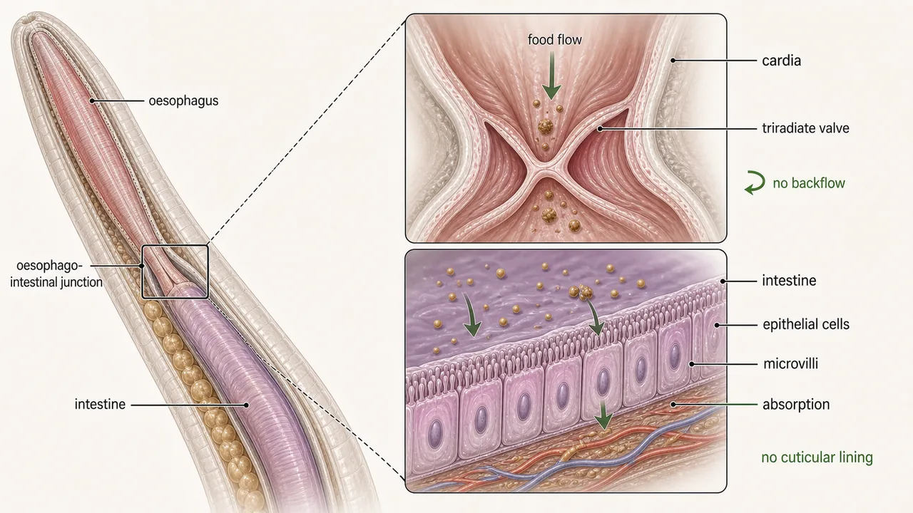
Mesenteron (Midgut) -- Intestine
The middle part of the digestive system is a hollow straight tube lined with a single layer of endodermal epithelial cells.
| Feature | Details |
|---|---|
| Cuticular lining (intima) | Absent (distinguishes midgut from cuticle-lined foregut and hindgut) |
| Microvilli | Finger-like projections of the plasma membrane into the lumen |
| Function of microvilli | Increase surface area for both secretion and absorption -- similar to villi in the human small intestine |
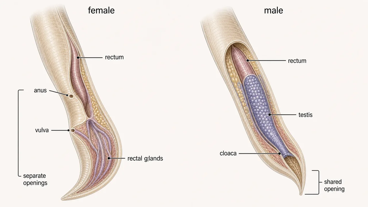
Proctodaeum (Hindgut) -- Rectum and Anus
The hindgut comprises the rectum and anus, regulating the expulsion of undigested food.
Male vs Female -- A Key Distinction
| Feature | Female | Male |
|---|---|---|
| Digestive opening | Anus (separate from reproductive opening) | Cloaca (shared with reproductive system) |
| Reproductive opening | Vulva (separate) | Through cloaca |
In male nematodes, the rectum joins with the hind part of the testis, forming a common opening called the cloaca (Latin for "common sewer"). In females, the anus and vulva are separate openings.
TIP
Key exam distinction: Males have a cloaca (shared opening for digestive and reproductive systems). Females have separate anus and vulva. This is a frequently tested difference.
Rectal glands are present in some species and secrete the gelatinous matrix in which eggs are deposited (e.g., the egg mass of Meloidogyne).
Summary Table
| Structure | Location | Key Feature | Exam Tip |
|---|---|---|---|
| Oral opening | Anterior end | Surrounded by 6 hexaradially arranged lips | Hexaradial symmetry |
| Stylet | Stoma | Hollow, needle-like; pierces cell walls | Stomatostylet (Secernentea) vs Odontostylet (Adenophorea) |
| Oesophagus | Between stoma and intestine | Muscular pump; prevents regurgitation | PPNs have 3-part oesophagus: corpus, isthmus, basal bulb |
| Oesophageal glands | Associated with oesophagus | Secrete enzymes into host cells | Create giant cells and syncytia |
| Cardia | Oesophago-intestinal junction | Triradiate valve prevents backflow | One-way valve |
| Intestine (midgut) | Central body | Microvilli for absorption; no cuticular lining | Secretory + absorptive function |
| Anus | Posterior (female) | Separate from vulva | Female only |
| Cloaca | Posterior (male) | Common opening for digestive + reproductive | Male only |
| Rectal glands | Hindgut | Secrete gelatinous egg mass matrix | Important in Meloidogyne |
TIP
Exam mnemonic -- "SIC" for foregut parts (mouth to intestine): Stoma (with stylet) --> Isthmus-containing oesophagus (3 parts) --> Cardia (triradiate valve).
References
- Walia, R. K and Bajaj, H. K (2014). Textbook of Introductory Plant Nematology. Directorate of Knowledge Management in Agriculture, ICAR, New Delhi.
- Ravichandra, N. G. (2019). Plant Nematology. I. K. International Publishing House Pvt. Ltd., New Delhi.
- Dasgupta, M. K. (1998). Phytonematology. Pilgrims Publishing
- Fotedar, D.N. & Handoo, Z.A. (1978) A revised scheme of classification to order Tylenchida Thorne, 1949 (Nematoda). Journal of Science, University of Kashmir (1975), 3, 55-82.
- Qing, X., Bert, W. Family Tylenchidae (Nematoda): an overview and perspectives. Org Divers Evol 19, 391-408 (2019).
- https://doi.org/10.1007/s13127-019-00404-4
- https://nematode.unl.edu/dolichod.htm
- http://www.nematologia.com.br/files/tematicos/6.pdf
Summary Cheat Sheet
| Concept / Topic | Key Details |
|---|---|
| Oral opening | Anterior end; Surrounded by 6 hexaradially arranged lips — Hexaradial symmetry |
| Stylet | Stoma; Hollow, needle-like; pierces cell walls — Stomatostylet (Secernentea) vs Odontostylet (Adenophorea) |
| Oesophagus | Between stoma and intestine; Muscular pump; prevents regurgitation — PPNs have 3-part oesophagus: corpus, isthmus, basal bulb |
| Oesophageal glands | Associated with oesophagus; Secrete enzymes into host cells — Create giant cells and syncytia |
| Cardia | Oesophago-intestinal junction; Triradiate valve prevents backflow — One-way valve |
| Intestine (midgut) | Central body; Microvilli for absorption; no cuticular lining — Secretory + absorptive function |
| Anus | Posterior (female); Separate from vulva — Female only |
| Cloaca | Posterior (male); Common opening for digestive + reproductive — Male only |
| Rectal glands | Hindgut; Secrete gelatinous egg mass matrix — Important in Meloidogyne |
TIP
Next: Lesson 06 covers physiology -- reproductive modes (sexual, parthenogenetic, hermaphroditic), excretory system types, and the nervous system.