⼍ Cell Coverings: Membrane, Wall, and Protoplasm
Understand plasma membrane structure, cell wall composition, protoplasm properties, and how these coverings protect plant cells — with agricultural examples and exam tips.
Why Cell Coverings Matter in Agriculture
Every time a farmer stores potatoes in a cold room or a food scientist processes sugarcane into jaggery, cell coverings play a central role. The cell wall gives sugarcane its rigidity, pectin in the middle lamella is what makes fruit jams set, and the plasma membrane controls which nutrients a root hair absorbs from the soil. Understanding these structures is essential for crop improvement, post-harvest technology, and disease resistance breeding.
Plasma Membrane (Cell Membrane / Plasmalemma)

The plasma membrane is the outermost living boundary of every cell. It is extremely thin, elastic, and selectively controls what enters and exits the cell. In direct cell-biology recall, it is also called the plasmalemma.
Structure (Fluid Mosaic Model)
- In plant cells, the membrane lies on the inner side of the cell wall.
- It is a lipoprotein membrane:
- Protein layer — 20 Å thickness
- Lipid bilayer — 35 Å
- Protein layer — 20 Å (monolayer)
- Total thickness: 75–100 Å
- In older exam-oriented one-liners, the plasma membrane is also described as a lipoprotein complex with roughly phospholipid 60% and protein 40% composition.
The Fluid Mosaic Model describes the membrane as a flexible, fluid lipid bilayer with proteins either embedded within it or attached to its surface.
Pro Content Locked
Upgrade to Pro to access this lesson and all other premium content.
Charged once for one year · ₹1188 total
Save ₹100/month vs ₹2388/year launch price
- All Agriculture & Banking Courses
- AI Lesson Questions (100/day)
- AI Doubt Solver (50/day)
- Glows & Grows Feedback (30/day)
- AI Section Quiz (20/day)
- 22-Language Translation (100/day)
- Recall Questions (20/day)
- AI Quiz (15/day)
- AI Quiz Paper Analysis (100/day)
- AI Step-by-Step Explanations (100/day)
- Spaced Repetition Recall (FSRS)
- AI Tutor
- Immersive Text Questions
- Audio Lessons — Hindi & English
- Mock Tests & Previous Year Papers
- Summary & Mind Maps
- XP, Levels, Leaderboard & Badges
- Generate New Classrooms
- Voice AI Teacher (AgriDots Live)
- AI Revision Assistant
- Knowledge Gap Analysis
- Interactive Revision (LangGraph)
🔒 Secure one-time yearly payment via Razorpay · No hidden fees
Why Cell Coverings Matter in Agriculture
Every time a farmer stores potatoes in a cold room or a food scientist processes sugarcane into jaggery, cell coverings play a central role. The cell wall gives sugarcane its rigidity, pectin in the middle lamella is what makes fruit jams set, and the plasma membrane controls which nutrients a root hair absorbs from the soil. Understanding these structures is essential for crop improvement, post-harvest technology, and disease resistance breeding.
Plasma Membrane (Cell Membrane / Plasmalemma)
The plasma membrane is the outermost living boundary of every cell. It is extremely thin, elastic, and selectively controls what enters and exits the cell. In direct cell-biology recall, it is also called the plasmalemma.
Structure (Fluid Mosaic Model)
- In plant cells, the membrane lies on the inner side of the cell wall.
- It is a lipoprotein membrane:
- Protein layer — 20 Å thickness
- Lipid bilayer — 35 Å
- Protein layer — 20 Å (monolayer)
- Total thickness: 75–100 Å
- In older exam-oriented one-liners, the plasma membrane is also described as a lipoprotein complex with roughly phospholipid 60% and protein 40% composition.
The Fluid Mosaic Model describes the membrane as a flexible, fluid lipid bilayer with proteins either embedded within it or attached to its surface.
- In classical membrane-model history, the older Sandwich Model is associated with Danielli and Davson, while the Unit Membrane Model is associated with David Robertson. These are mainly of historical exam importance, whereas the Fluid Mosaic Model of Singer and Nicolson remains the most accepted explanation.
Properties and Functions
| Property | Significance |
|---|---|
| Selectively permeable | Allows only certain molecules to pass, maintaining homeostasis. This is how root cells absorb water and nutrients while blocking harmful substances. |
| Ectoplast (in animals) | Since animal cells lack a cell wall, the plasma membrane is the only outer boundary. |
| Absent in viruses | Viruses have a protein coat (capsid) instead. |
| Contains sialic acid | A nine-carbon monosaccharide that acts as a cell receptor for cell recognition and signalling. |
Membrane Classification of Organelles
IMPORTANT
This classification is frequently tested in IBPS AFO and NABARD Grade A.
| Category | Organelles |
|---|---|
| Membrane-less | Ribosome, Centriole, Centrosome, Microtubules |
| Single membrane-bound | Peroxisomes, Lysosomes, Sphaerosome, Glyoxysomes |
| Double membrane-bound | Nucleus, Mitochondria, Chloroplast |
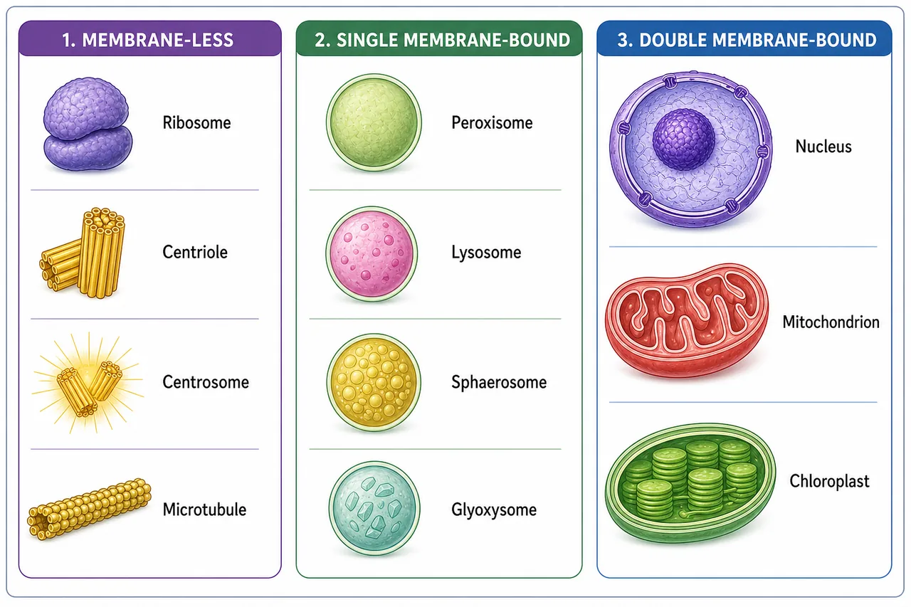
Mnemonic: Double-membrane = "NMC" (Nucleus, Mitochondria, Chloroplast). Both mitochondria and chloroplasts are semi-autonomous because they contain their own DNA.
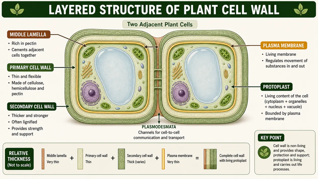
Cell Wall
NOTE
Cell wall is present in plant cells but absent in animal cells — this is one of the most fundamental differences tested in exams.
- The cell wall is a non-living, thick, rigid envelope outside the plasma membrane.
- It is permeable and made of cellulose — a long-chain polymer of glucose that gives tensile strength.
- Cell wall is absent in animal cells, which is why animal cells are more flexible in shape.
Agricultural connection: The toughness of jute and cotton fibres comes from the thick cellulose walls of their cells. Breeding for fibre quality in these crops directly involves selecting for cell wall properties.
Middle Lamella
The middle lamella is the cementing layer that joins the primary cell walls of adjacent cells, holding them together to form tissues.
- Rich in pectin (a polysaccharide used as cementing material and in jelly making).
- Classical exam books often describe the middle lamella as the outermost layer of the plant cell wall because it lies between two adjacent cells and cements them together.
- Contains Ca2+ and Mg2+, so older one-liners may summarise it as Ca-Mg pectate; functionally, this is the same cementing pectin-rich layer linking neighbouring cells.
Agricultural example: When fruits ripen, enzymes break down pectin in the middle lamella, causing the fruit to soften. This is why overripe mangoes become mushy — the cells literally come apart.
Primary Cell Wall
| Component | Chemical Nature | Role |
|---|---|---|
| Cellulose | Polymer of hexose (6C) glucose | Structural strength |
| Hemicellulose | Polymer of mannose/galactose/pentose | Matrix support |
| Pectin | Hydrophilic polysaccharide | Flexibility and water retention |
The primary wall is relatively thin and flexible, allowing cell expansion during growth. Pectin is in the primary cell wall in lower plants but in the middle lamella in higher plants.
- Much of the wall matrix material, especially pectins and hemicelluloses, is processed through the Golgi apparatus before being delivered to the developing cell wall.
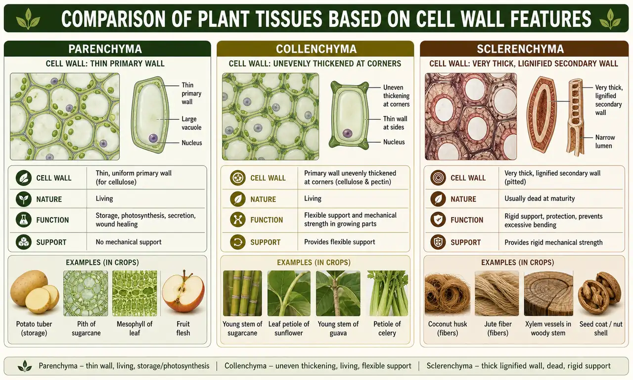
Plant Cell Types Based on Cell Wall
| Cell Type | Wall Composition | Properties | Examples |
|---|---|---|---|
| Parenchyma | Primary wall only (thin) | Soft, flexible; involved in photosynthesis, storage, repair | Mesophyll cells, potato tuber storage cells |
| Collenchyma | Primary + secondary wall (cellulose only) | Unevenly thickened; flexible mechanical support | Young stems, leaf stalks (petioles) |
| Sclerenchyma | Primary + thick secondary wall (cellulose + lignin) | Extremely rigid; usually dead at maturity | Fibres (jute, hemp), vessels, tracheids |
- In textbook one-liners, the secondary wall is absent in meristematic cells because actively dividing cells need expandable primary walls rather than rigid lignified reinforcement.
- In compact revision language, the secondary wall is the more rigid inner reinforcement layer and may accumulate extra materials such as lignin and, in specialized cells, substances like suberin that strengthen or waterproof the wall.
Lignin
- Chemically it is coniferyl alcohol, solid at room temperature.
- Hardness of woody tissue is due to lignin. Exams
- Lignin is the second most abundant organic polymer on Earth (after cellulose). It makes cell walls waterproof and structurally rigid.
| Cell Category | Lignin Status | Examples | Staining |
|---|---|---|---|
| Non-lignified | Absent | Parenchyma, collenchyma, sieve tubes | — |
| Lignified | Present | Sclerenchyma, vessels, tracheids | Safranin (red dye) |

Agricultural relevance: Lignin content determines the digestibility of crop residues used as animal fodder. Low-lignin sorghum varieties (brown midrib mutants) are bred specifically for better fodder quality.
Protoplasm
Protoplasm is all the living content of a cell including the plasma membrane — the cytoplasm, nucleus, and all organelles together.
-
In direct plant-cell exam language, Plant cell = Cell wall + Protoplast.
-
The protoplast is the living part of the cell bounded by the plasma membrane, so it can be recalled as Cell - Cell wall.
-
In classical cell-biology one-liners, the term cytoplasm is commonly credited to Kolliker / Strasburger; exam books often treat this as a direct recall fact alongside Purkinje's term protoplasm.
-
Older exam-oriented notes also associate the early recognition / discovery of protoplasm with Dujardin (1835).
| Property | Detail |
|---|---|
| Term coined by | J.E. Purkinje (1830–37) |
| Huxley's definition | "Physical basis of life" |
| Water content | 80–90% |
| Most abundant dry constituent | Protein (60–70%) |
| Nature | Polyphasic colloidal system (sol ↔ gel) |
Exam tip: "Protoplasm = physical basis of life" (Huxley) is one of the most frequently asked one-liners.
Nucleus
- Word derived from Latin "Kernel" — the central, essential core of the cell.
- Discovered by Robert Brown (1833) while studying orchid cells.
- In direct exam wording, the nucleus is often described as the storehouse of genetic material because it contains the chromosomes and genes that control heredity and cell activity.
- Surrounded by a double-membrane nuclear envelope (lipoprotein) with nuclear pores.
- Size: 5–25 µm.
Where Is the Nucleus Absent?
| Condition | Reason |
|---|---|
| Bacteria and cyanobacteria | Prokaryotes — DNA exists as nucleoid (no membrane-bound nucleus) |
| Mature mammalian RBCs | Nucleus lost to make room for haemoglobin |
| Sieve tube cells (phloem) | Lose nucleus at maturity; depend on companion cells |
| Xylem vessels/tracheids | Dead at maturity; hollow tubes for water transport |
Agricultural connection: Phloem sieve tubes transport sucrose from leaves to developing grains in wheat and rice. Their unique nucleus-free structure allows maximum flow of assimilates.
Nuclear Contents
- Nucleoplasm (nuclear sap/karyolymph) — gel-like matrix supporting chromatin and nucleolus.
- Chromatin — tangled thread-like mass of DNA + histone protein; condenses into chromosomes during cell division.
- Genes — stretches of DNA that carry information for protein synthesis. Genes are the hereditary units; DNA is the hereditary material.
Nucleolus
- A spheroidal, non-membrane-bound, dense organelle within the nucleus.
- Discovered by Fontana (1781). Rich in RNA but also contains DNA.
- Main function: synthesise ribosomal RNA (rRNA).
- Attached to a specific chromosomal site called the nucleolar organiser region (NOR).
RNA Polymerases in Eukaryotes
| Enzyme | Location | Product |
|---|---|---|
| RNA Polymerase I (A) | Nucleolus | rRNA |
| RNA Polymerase II (B) | Nucleoplasm | HnRNA (precursor of mRNA) |
| RNA Polymerase III (C) | Nucleoplasm | tRNA (sRNA) |
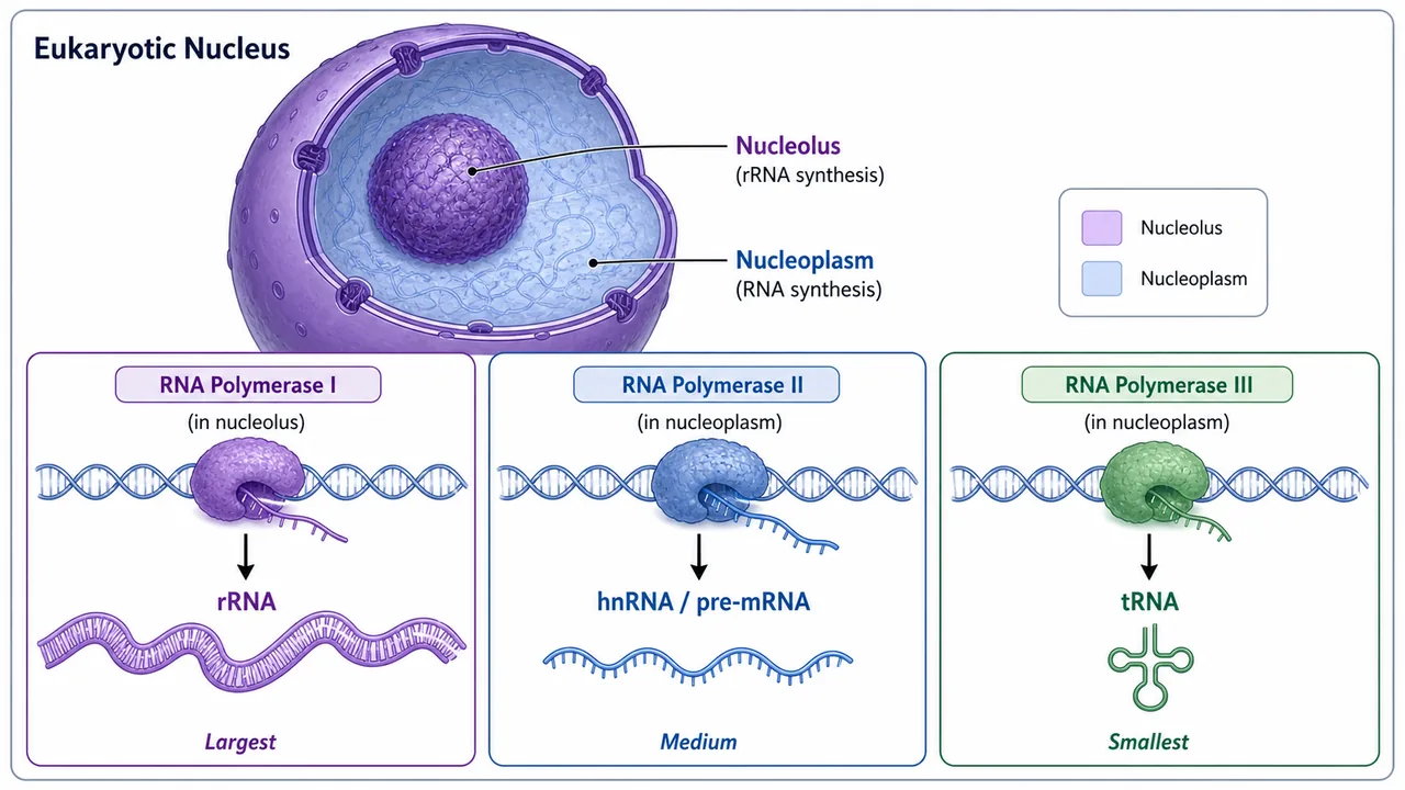
Exam tip: Remember "I = ribosomal, II = messenger, III = transfer" — the numbering matches the size of product (rRNA is largest, tRNA is smallest).
Chromosome

- First seen by Strasburger in 1875 as fine threads.
- Named "Chromosome" (chroma = colour + soma = body) by Waldeyer in 1888 using basic dye.
- Chromosomes are carriers of hereditary units (genes) — established by Morgan using Drosophila. UPPSC 2021
- Homozygous = identical alleles for a trait; Heterozygous = different alleles. Exams

- Chromosome number is species-specific: human 2n = 46, rice 2n = 24.
- In prokaryotes: single circular chromosome called genophore (without histones).
- In eukaryotes: rod-shaped, linear chromosomes wrapped around histone proteins.

Genetic Material: DNA and RNA
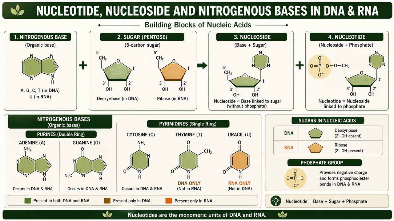
Building Blocks
| Term | Composition |
|---|---|
| Nucleotide | Sugar + Nitrogenous base + Phosphate (H₃PO₄) |
| Nucleoside | Sugar + Base only |
| Relationship | Nucleotide = Nucleoside + Phosphoric acid |
Nitrogenous Bases
| Base | Type | Found in |
|---|---|---|
| Adenine (A) | Purine (double ring) | DNA and RNA |
| Guanine (G) | Purine (double ring) | DNA and RNA |
| Cytosine (C) | Pyrimidine (single ring) | DNA and RNA |
| Thymine (T) | Pyrimidine (single ring) | DNA only |
| Uracil (U) | Pyrimidine (single ring) | RNA only |
Mnemonic: Pure As Gold = Purines are Adenine and Guanine. The rest (C, T, U) are pyrimidines.
Key Discoveries
| Scientist | Year | Contribution |
|---|---|---|
| Miescher | 1868 | First isolated nucleic acids ("nuclein") from WBC pus |
| Avery, MacLeod & McCarty | 1944 | Proved DNA (not protein) is the genetic material |
| Watson & Crick | 1953 | Double Helix Model of DNA |
| Wilkins & Franklin | — | X-ray diffraction data of DNA |
| Watson, Crick & Wilkins | 1962 | Nobel Prize for DNA structure |
| A. Kornberg | — | First in vitro synthesis of DNA |
| S. Ochoa | — | In vitro synthesis of RNA |
| H.G. Khorana & K.L. Agrawal | — | Artificial synthesis of alanine tRNA gene |
Structure of DNA (Watson-Crick Model)
- Two antiparallel polynucleotide chains wound helically (one runs 5'→3', the other 3'→5').
- Sugar-phosphate backbone on the outside; bases on the inside.
- Chargaff's Rules: A = T and G = C; total purines (A+G) = total pyrimidines (T+C).
- (A+T)/(G+C) = Base pair ratio — unique to each species.
| Parameter | Value |
|---|---|
| Base pairs per turn | 10 |
| Distance between base pairs | 3.4 Å |
| Length per turn | 34 Å |
| Diameter of helix | 20 Å |
| A–T bonds | 2 hydrogen bonds |
| G–C bonds | 3 hydrogen bonds (more stable) |

Exam tip: Higher G-C content = higher thermal stability (melting temperature). This matters when designing PCR primers in agricultural biotechnology.
Structure of RNA
- Usually single-stranded (not helical like DNA).
- In most plant viruses, genetic material is RNA.
- Non-genetic RNAs: mRNA, tRNA, rRNA — synthesised on DNA template.
- Genetic RNA of viruses is self-replicating via RNA-dependent RNA synthesis using the enzyme RdRp.

| Virus | Type of RNA |
|---|---|
| Plant Viruses | |
| Turnip yellow mosaic virus (TYMC) | Single stranded |
| Wound tumour | Double stranded |
| Animal viruses | |
| Influenza virus | Single stranded |
| Rous Sarcoma | Single stranded |
| Poliomyelitis | Single stranded |
| Reovirus | Double stranded |
| Bacteriophages | |
| MS 2, F 2, r 17 | Single stranded |
Gene Concepts
| Concept | Scientist | Key Idea |
|---|---|---|
| One gene–one enzyme | Beadle & Tatum (1943) on Neurospora crassa | Each gene codes for one enzyme |
| Operon concept | Jacob & Monod | How gene expression is regulated (lac operon) |
| Gene fine structure | Benzer | Genes can be divided into functional sub-units |
Benzer's three units:
| Unit | Definition | Size |
|---|---|---|
| Recon | Smallest unit of recombination | 1–2 nucleotide pairs (smallest) |
| Muton | Smallest unit of mutation | Single nucleotide pair |
| Cistron | Functional unit (= gene in practice) | Hundreds of nucleotide pairs (largest) |
Modern refinement: One gene → One polypeptide (not all genes code for enzymes, and some proteins have multiple polypeptide chains).
Genetic Code
- Deciphered by Holley, Khorana & Nirenberg (Nobel Prize 1968).
- Triplet code: 4 bases taken 3 at a time = 4³ = 64 codons (enough for 20 amino acids).
| Property | Meaning |
|---|---|
| Degenerate | More than one codon can code for the same amino acid (e.g., Arg, Ser, Leu each have 6 codons) |
| Non-overlapping | Each base belongs to only one codon |
| Comma-less | No punctuation between codons; reading is continuous |
| Universal | Same code in all organisms (minor exceptions in mitochondrial DNA) |
| Ambiguous | Under abnormal conditions (e.g., streptomycin), a codon may code for a different amino acid |
| Codon Type | Codons | Function |
|---|---|---|
| Start codon | AUG | Initiates translation; codes for methionine |
| Stop codons | UAA (ochre), UAG (amber), UGA (opal) | Terminate translation; do not code for any amino acid |

Mnemonic for stop codons: "U Are Annoying, U Are Gone, U Go Away" — UAA, UAG, UGA.
Mitochondria
TIP
Organelle nicknames: Mitochondria = "Power house", Chloroplast = "Kitchen of the cell", Lysosome = "Suicidal bag", Golgi = "Post office".
- Power house of the cell — primary site of ATP production through aerobic respiration.
- ATP = Energy currency of the cell.
- First identified by Altman (1886) as Bioplast.
- Named "mitochondria" by C. Benda (1898).

| Process | Location |
|---|---|
| Glycolysis | Hyaloplasm (cytosol), NOT mitochondria |
| Krebs cycle (aerobic respiration) | Mitochondrial matrix |
| Electron transport chain + ATP synthesis | Inner membrane (oxysomes/F1 particles) |
- Contains own DNA (0.02%), RNA (3–4%), and 70S ribosomes → semi-autonomous (supports Endosymbiotic Theory).
Plastids
Classified by Schimper (1885) based on pigment content:
| Plastid Type | Colour | Function | Agricultural Example |
|---|---|---|---|
| Chloroplast | Green | Photosynthesis | Leaf mesophyll of all green crops |
| Chromoplast | Red/Yellow/Orange | Attract pollinators and seed dispersers | Tomato, carrot, marigold flowers |
| Leucoplast | Colourless | Food storage | Potato tubers, cereal grains |
Types of Leucoplasts
| Type | Stores | Example |
|---|---|---|
| Amyloplast | Starch | Potato tubers, rice grains |
| Elaioplast | Oils | Groundnut, mustard seeds |
| Aleuronoplast | Protein | Aleurone layer of wheat/rice |
Chlorophyll and Plant Pigments
| Pigment | Colour | Formula | % in Green Plants |
|---|---|---|---|
| Chlorophyll a | Blue-black | C₅₅H₇₂O₅N₄Mg | 65% (Chl a + b combined) |
| Chlorophyll b | Green-black | C₅₅H₇₀O₆N₄Mg | |
| Xanthophyll | Yellow | C₄₀H₅₆O₂ | 29% |
| Carotene | Yellowish-orange | C₄₀H₅₆ | 6% |
- Carotene + Xanthophyll = carotenoid pigments (accessory pigments; provide photoprotection).
- Chromoplasts contain only carotenoid pigments.
Pigment Classification
| Category | Solubility | Location | Examples |
|---|---|---|---|
| Plastid pigments | Organic solvents only (lipid-soluble) | Plastid membranes | Chlorophyll, carotenoids |
| Sap pigments | Water-soluble | Vacuoles | Anthocyanin (red/purple/blue in flowers and beets) |
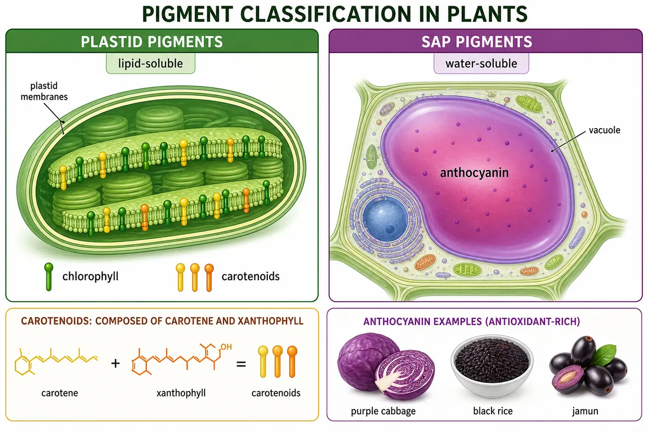
Agricultural note: Anthocyanin content in crops like purple cabbage, black rice, and jamun is a marker for antioxidant-rich varieties — an active area of breeding research.
Chloroplast Structure

| Region | Key Structures | Reaction |
|---|---|---|
| Grana (thylakoid stacks) | Quantasomes with chlorophyll | Light reactions → ATP + NADPH |
| Stroma (matrix) | Enzymes including RuBISCO | Dark reactions (Calvin cycle) → sugar |
- RuBISCO is the most abundant protein on Earth.
- Chloroplasts contain own DNA (0.5%), RNA (3–4%), and 70S ribosomes → semi-autonomous (Endosymbiotic Theory).
- Grana are interconnected by stroma lamellae (intergranal lamellae).
Endoplasmic Reticulum (ER)

- Dense network of double-membrane structures running through the cytoplasm.
- Ultrastructure first reported by Porter (1948). Origin from nuclear membranes.
- Dynamic — can be broken down and reconstructed based on cellular needs.
| Type | Feature | Primary Function |
|---|---|---|
| Rough ER (RER) | Ribosomes attached | Protein synthesis (for secretion/membranes) |
| Smooth ER (SER) | No ribosomes | Lipid synthesis, steroid production, detoxification |
Functions of ER
- Provides mechanical support (endoskeleton) to the cell.
- Increases surface area for metabolic reactions.
- Intracellular transport of molecules.
- Formation of cell plate and nuclear membrane during division.
- SER produces lipids for membrane biogenesis.
- SER in liver cells detoxifies drugs and poisons.
Ribosomes
- Composition: rRNA (40–60%) + Protein (40–60%) — no lipid (membrane-less organelle).
- First observed by Claude (1943); term coined by R.B. Robert (1958).
- Detailed ultrastructure by Palade (1956, Nobel Prize).
| Organism Type | Ribosome Size | Subunits |
|---|---|---|
| Prokaryotes & chloroplasts | 70S | 50S + 30S |
| Eukaryotes (cytoplasm) | 80S | 60S + 40S |
- Mg²⁺ ions promote subunit association; low Mg²⁺ causes dissociation.
Types of RNA
| RNA Type | % of Total | Function |
|---|---|---|
| mRNA (messenger) | 5–10% | Carries genetic instructions from DNA to ribosomes |
| tRNA (transfer/sRNA) | 10–15% | Carries amino acids; clover-leaf shape; smallest |
| rRNA (ribosomal) | 80% | Structural/catalytic core of ribosomes; most stable |
Protein Synthesis Steps
- Transcription — DNA → mRNA (in nucleus)
- Translation — mRNA → Protein (at ribosomes)


Explore More
Golgi Body (Dictyosome)
- Discovered by Camillo Golgi (1898) using silver staining in nerve cells.
- Stacks of flattened sacs called cisternae; called dictyosomes in plants.
- Has polarity: cis face (receiving, near ER) → trans face (shipping, near plasma membrane).
- Origin from ER (transition vesicles).
- Acrosomes on sperm cells are derived from Golgi complex.

Functions
- Store, modify, package and condense proteins (the cell's "post office").
- Form the cell plate during plant cell division.
- Add sugars to proteins (glycosylation → glycoproteins).
- Produce lysosomes.

Lysosome

- Lysis = digestion; Soma = body → "digestive bodies" of the cell.
- Single membrane-bound vesicles containing hydrolytic enzymes.
- Formed from Golgi complex (directly) and ER (indirectly).
- Discovered by De Duve (1955, Nobel Prize 1974). Mainly found in animals; also in some plants like Neurospora.
Functions
- Intracellular digestion of large molecules.
- Defense against bacteria and viruses (fuse with phagosomes).
- During starvation: digest own organelles (autophagy) → called "suicidal bag".
Other Organelles and Structures
Spherosomes
- Single membrane-bound; mainly in plants.
- Function: fat metabolism — abundant in oilseeds (groundnut, mustard, sunflower).
Microsomes
- Artificial structures formed when cells are broken in the lab (ER fragments + ribosomes).
- Used as in vitro models to study ER functions and protein synthesis.
Vacuole

- Most prominent in mature plant cells; may occupy up to 90% of cell volume.
- Single membrane-bound (membrane = tonoplast); contains cell sap.
- Functions: osmoregulation, storage, maintaining turgor pressure (cell rigidity).
Agricultural relevance: Turgor pressure keeps crop plants upright. When water supply drops, cells lose turgor and the plant wilts — visible as leaf rolling in rice or drooping in sunflower.
Plasmodesmata

- Microscopic channels found only in plants; named by Strasburger (1903).
- Origin from ER.
- Allow direct cell-to-cell communication and transport of molecules.
- All connected protoplasts form the symplast.
Centrosome
- Present near nucleus in all animal cells and some plant groups (Chlamydomonas, gymnosperms).
- Contains two centrioles (nine triplets of microtubules, 9+0 arrangement).
- Functions as the microtubule organising centre (MTOC); produces astral rays during mitosis.
Ergastic Substances
- Non-living cell inclusions: starch, sugar, organic acids, fats, oils, pigments, crystals, tannins, resins.
- These are metabolic products, not part of living protoplasm.
Summary Table
| Topic | Key Fact | Exam Pointer |
|---|---|---|
| Plasma membrane | Lipoprotein, 75–100 Å thick | Fluid Mosaic Model |
| Cell wall | Cellulose (non-living, permeable) | Present in plants, absent in animals |
| Middle lamella | Pectin + calcium pectate | Cementing layer; pectin used in jelly making |
| Lignin | Coniferyl alcohol | Hardness of wood; stained by Safranin |
| Protoplasm | Physical basis of life (Huxley) | 80–90% water; protein most abundant dry component |
| Nucleus | Discovered by Robert Brown (1833) | Absent in mature RBCs, sieve tubes, xylem |
| Nucleolus | Synthesises rRNA | Attached to NOR on chromosome |
| Chromosome | Named by Waldeyer (1888) | Species-specific number; carriers of genes (Morgan) |
| DNA | Double helix (Watson & Crick, 1953) | Chargaff's rule: A=T, G=C |
| Genetic code | Triplet, degenerate, universal | AUG = start; UAA, UAG, UGA = stop |
| Mitochondria | Power house; Krebs cycle in matrix | Semi-autonomous; glycolysis in hyaloplasm |
| Chloroplast | Kitchen of cell; light rxn in grana | Semi-autonomous; RuBISCO in stroma |
| Ribosome | 70S (prokaryotes) / 80S (eukaryotes) | No lipid; membrane-less |
| Golgi body | Post office of cell | Forms cell plate and lysosomes |
| Lysosome | Suicidal bag (De Duve, 1955) | Autophagy during starvation |
| Vacuole | Up to 90% of plant cell volume | Tonoplast = vacuolar membrane |
Summary Cheat Sheet
| Concept / Topic | Key Details |
|---|---|
| Plasma membrane | 75–100 Å thick; Fluid Mosaic Model; lipoprotein |
| Cell wall | Non-living, rigid, cellulose-based; permeable; absent in animal cells |
| Middle lamella | Cementing layer; rich in pectin + calcium pectate |
| Lignin | Coniferyl alcohol; hardness of wood; stained by Safranin |
| Parenchyma | Primary wall only; soft, flexible (photosynthesis, storage) |
| Collenchyma | Unevenly thickened wall; flexible support (young stems) |
| Sclerenchyma | Thick wall with lignin; dead at maturity (jute, hemp fibres) |
| Protoplasm coined by | J.E. Purkinje (1830–37); "Physical basis of life" = Huxley |
| Protoplasm composition | 80–90% water; protein = most abundant dry component |
| Nucleus discovered by | Robert Brown (1833) — in orchid cells |
| Nucleus absent in | Bacteria, mature RBCs, sieve tubes, xylem vessels |
| Nucleolus | Synthesises rRNA; attached to NOR on chromosome |
| RNA Pol I = rRNA | RNA Pol II = mRNA; RNA Pol III = tRNA |
| Chromosome named by | Waldeyer (1888); first seen by Strasburger (1875) |
| DNA double helix | Watson & Crick (1953); A=T (2 H-bonds), G=C (3 H-bonds) |
| DNA parameters | 10 bp/turn, 3.4 Å between bases, 34 Å/turn, 20 Å diameter |
| Genetic code | Triplet, degenerate, universal; AUG = start; UAA/UAG/UGA = stop |
| Benzer's units | Recon (recombination) < Muton (mutation) < Cistron (function) |
| One gene–one enzyme | Beadle & Tatum (1943) on Neurospora crassa |
| Mitochondria | "Power house"; named by C. Benda (1898); Krebs cycle in matrix |
| Glycolysis occurs in | Hyaloplasm (cytosol), NOT mitochondria |
| Chloroplast | Light reactions in grana; dark reactions in stroma |
| RuBISCO | Most abundant protein on Earth; in stroma |
| Chlorophyll a | 65% of pigments; blue-black; formula C₅₅H₇₂O₅N₄Mg |
| Anthocyanin | Water-soluble sap pigment in vacuoles |
| Ribosome sizes | 70S (prokaryotes) = 50S+30S; 80S (eukaryotes) = 60S+40S |
| Golgi body | "Post office"; discovered by Camillo Golgi (1898) |
| Lysosome | "Suicidal bag"; De Duve (1955); autophagy during starvation |
| Vacuole | Up to 90% of plant cell; membrane = tonoplast |
| Spherosomes | Fat metabolism; abundant in oilseeds |
| Plasmodesmata | Plant cell-to-cell channels; form symplast |