🐞 Insect Exoskeleton and Moulting
Structure and functions of the insect integument, layers of the cuticle, cuticular modifications, and the hormonal control of the moulting process
In the previous unit, we classified Phylum Arthropoda into seven classes and identified the defining features of Class Insecta. Now we begin the study of insect external morphology -- starting with the body wall that makes terrestrial life possible.
When a farmer sprays contact insecticide on a pest, the first barrier the chemical must cross is the insect's exoskeleton -- particularly the waxy epicuticle. Understanding the structure of this body wall explains why some insecticides work on contact (they dissolve the wax layer) while others must be ingested. The exoskeleton also determines when insects are most vulnerable -- just after moulting, when the new cuticle is still soft and unprotected.
This lesson covers:
- Insect morphology -- what it is and why it matters for pest identification
- Integument structure -- three layers (cuticle, epidermis, basement membrane) and their functions
- Cuticular modifications -- external outgrowths and internal invaginations
- Moulting (ecdysis) -- the three-stage process and the hormones that control it
What Is Insect Morphology?
- The branch of science dealing with the study of physical structure and form of insects.
- It covers the head, thorax, abdomen, legs, wings, and mouthparts.
- Morphological knowledge is essential for accurate pest identification and understanding how body structures relate to insect behaviour and ecology.
- The arrangement of body bristles or setae is called chaetotaxy, an especially useful idea in larval identification and external morphology.
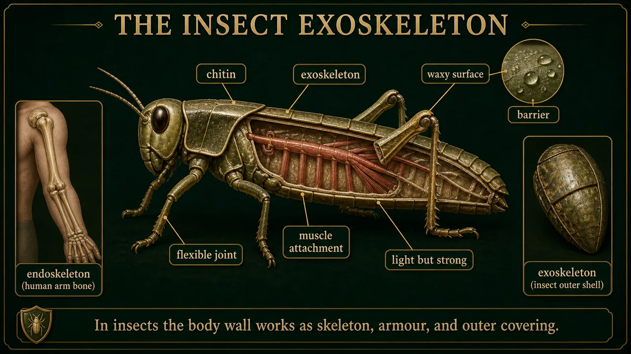
The Insect Exoskeleton
Think of it this way: Humans have bones inside their body (endoskeleton). Insects wear their skeleton on the outside -- like a suit of armour that also serves as skin, raincoat, and muscle anchor all in one. This "armour-skin" is the exoskeleton.
- The external covering or body wall is made of a hard polymer called chitin -- a nitrogen-containing polysaccharide that provides remarkable strength while remaining lightweight. Think of chitin as nature's plastic -- tough, light, and mouldable.
- This outer covering acts as the skeletal system and is called the exoskeleton (unlike the internal endoskeleton of vertebrates).
- The insect body wall is also called the integument (Latin: "covering").
- It is rigid yet flexible, lighter than bone, and variously modified in different body parts -- thicker on the head and thorax for protection, thinner at joints for movement (just as a knight's armour has plates at the chest but flexible chainmail at the elbows).
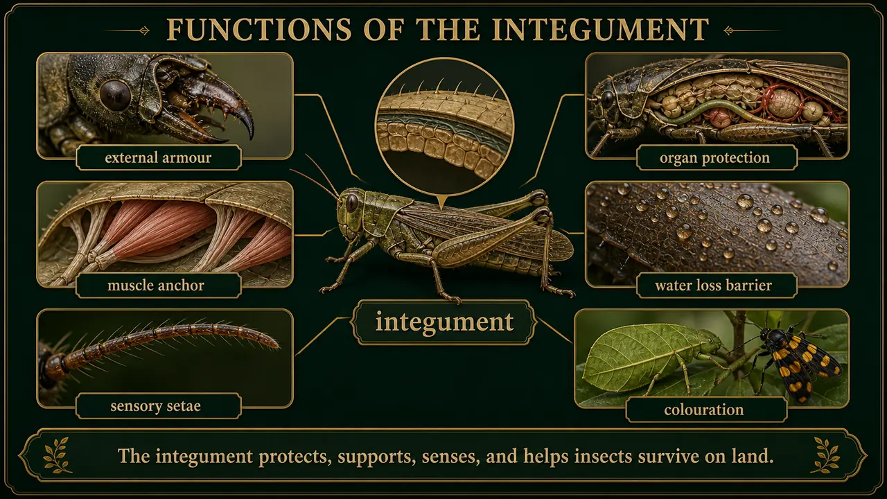
Functions of the Integument
| Function | How It Works |
|---|---|
| External armour | Protects external organs (jaws, ovipositor) from physical damage |
| Internal organ protection | Shields foregut, hindgut, trachea from chemicals, parasites, predators, pathogens |
| Muscle attachment & body shape | Provides anchor points for muscles and maintains body form |
| Prevents water loss | Reduces evaporation -- critical for terrestrial insects |
| Sensory detection | Contains sensory structures that detect touch, chemicals, temperature |
| Colouration | Contains pigments for camouflage, warning colouration, or mate attraction |
Structure of the Integument
The insect integument consists of three layers, from outside to inside -- imagine peeling an onion, each layer has a distinct job:
Building analogy: The integument is like a painted wall. The cuticle is the paint and plaster (non-living, protective), the epidermis is the bricklayer who built it (living, active), and the basement membrane is the foundation slab (non-living, structural support).
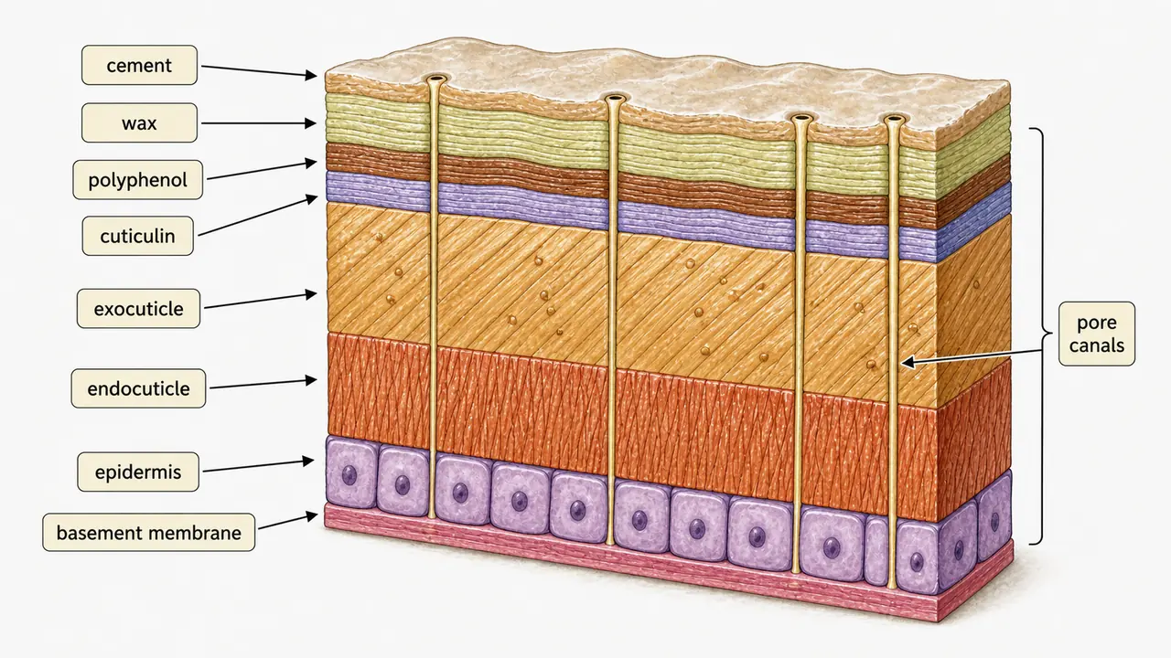
1. Cuticle (outermost, non-living)
- Non-cellular layer secreted by the epidermis.
- Made of chitin (35--60%) and protein (25--40%).
- Chitin is a nitrogenous polysaccharide made of N-acetylglucosamine chains -- probably the second most abundant polysaccharide in nature after cellulose.
- Divided into epicuticle (non-chitinous, thin) and procuticle (chitinous, thick).
- The chitinous procuticle later differentiates into exocuticle and endocuticle.
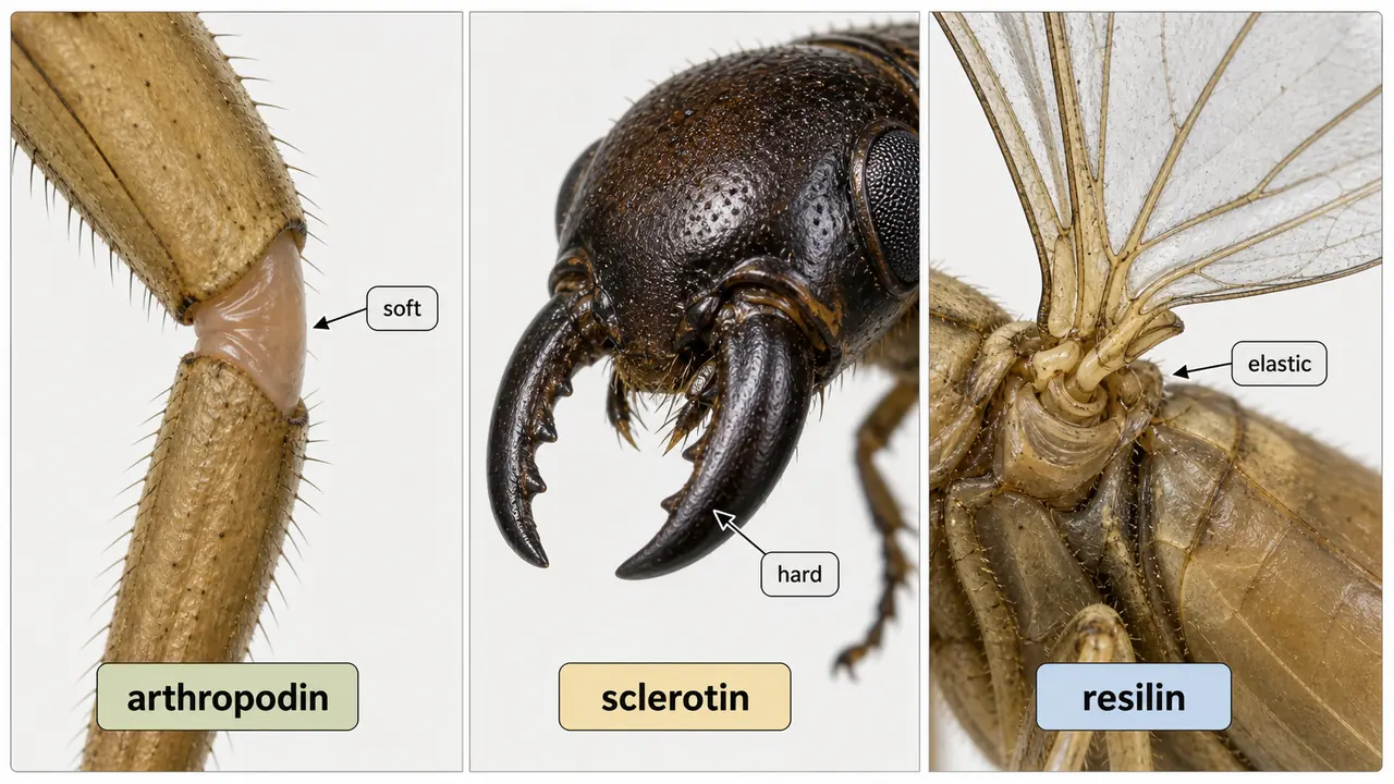
Key Proteins in the Cuticle
Everyday comparison: These three proteins are like three building materials -- Arthropodin is like raw clay (soft, flexible, easily washed away), Sclerotin is like fired brick (hard, permanent, dark), and Resilin is like a rubber band (stretches and snaps back). Each is used where its properties are needed most.
| Protein | Nature | Property | Location |
|---|---|---|---|
| Arthropodin | Untanned, water-soluble | Soft, flexible | Soft body regions, joints |
| Sclerotin | Tanned (cross-linked), water-insoluble | Hard, rigid, dark-coloured | Sclerotized regions (head capsule, mandibles) |
| Resilin | Elastic | Stores and releases energy like rubber | Wing hinges, leg joints |
A. Epicuticle (outermost, non-chitinous)
Very thin (1--4 microns -- thinner than a human hair) but serves as the insect's first line of defence against water loss and chemicals. Contains no chitin. Think of epicuticle as a waterproof raincoat worn over the main armour -- without it, the insect would dry up and die within hours.
Consists of four sub-layers (outside to inside):
| Layer | Composition | Function |
|---|---|---|
| Cement layer | Lipoprotein (secreted by dermal glands) | Protects wax layer from abrasion |
| Wax layer | Long-chain hydrocarbons, fatty acid esters (0.25 microns) | Waterproofing -- prevents desiccation; the most critical layer for terrestrial survival |
| Polyphenol layer | Phenolic compounds | Contributes to hardening and darkening; resistant to acids and solvents |
| Cuticulin layer | Amber-coloured thin layer | Controls permeability; acts as growth barrier; first layer deposited during new cuticle formation |
Agricultural connection: Contact insecticides like diatomaceous earth and some oils work by disrupting the wax layer, causing the insect to lose water and die from desiccation.
Sequence recall: During new cuticle formation, the cuticulin layer is deposited first, followed by the other epicuticular and procuticular layers.
B. Procuticle (inner, chitinous)
Forms the structural bulk of the cuticle. After sclerotization, it differentiates into:
| Sub-layer | Colour | Hardness | Key Protein | Digested during moulting? |
|---|---|---|---|---|
| Exocuticle | Darkly pigmented | Hard, sclerotized | Sclerotin | No -- shed as part of old cuticle |
| Endocuticle | Light-coloured | Soft, unsclerotized | Arthropodin | Yes -- partially digested and recycled |
Pore Canals
- Fine vertical channels (0.1--0.15 microns diameter) running through both exo- and endocuticle.
- Act as microscopic highways transporting cuticular material and enzymes from the epidermis to the outer cuticle.
2. Epidermis / Hypodermis (middle, the only living layer)
- A unicellular layer of active glandular cells.
- Main function: secretes the cuticle above it.
- Also digests and absorbs old cuticle during moulting, and repairs wounds.
- Specialised cell types include:
- Dermal glands -- produce cement layer
- Trichogen cells -- produce hair-like setae
- Moulting glands -- secrete moulting fluid
- Peristigmatic glands -- around spiracles (in Dipteran larvae)
3. Basement Membrane (innermost, non-living)
- Non-cellular, amorphous granular layer formed from degenerated epidermal cells.
- Provides the foundation for the integument; muscles attach to it.
Cuticular Modifications
The cuticle is modified into external outgrowths and internal invaginations that greatly increase its functional versatility.
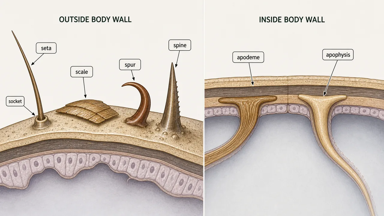
A. External Outgrowths
I. Cuticular Appendages (with membranous joint -- movable)
Setae (Macrotrichia):
- Commonly called hairs; arise from a cup-like pit (alveolus).
- Unicellular; produced by a trichogen cell; socket formed by a tormogen cell.
- Study of setal arrangement = chaetotaxy (useful for identifying larval stages).
| Type of Seta | Description | Example |
|---|---|---|
| Clothing hairs / Bristles | Hair-like structures covering body, legs, wings | Honeybee, flies |
| Scales | Pigmented plate-like structures (flattened modified setae / body-wall outgrowths) | Moths, butterflies (Lepidoptera) |
| Glandular setae | Outlet for hypodermal gland secretions | Certain Lepidoptera larvae |
| Sensory setae | Equipped with sensory receptors for touch, chemicals | Antennae, legs, mouthparts |
Spurs: Found on legs; multicellular in origin; stouter than setae; often at tips of tibia.
II. Cuticular Processes (no membranous joint -- fixed)
| Type | Description | Example |
|---|---|---|
| Microtrichia (fixed hairs / aculei) | Minute hair-like structures | Wings of Mecoptera, certain Diptera |
| Spines | Thorn-like, immovable extensions | Often defensive in function |
B. Internal Invaginations
| Type | Structure | Function |
|---|---|---|
| Apodemes | Hollow cuticular invaginations | Muscle attachment (like internal "shelves") |
| Apophyses | Solid cuticular invaginations | Mechanical support for organs |
Moulting (Ecdysis)
Moulting is the periodic shedding of the old cuticle accompanied by the formation of a new cuticle. It is the only way insects can grow larger, since the hard exoskeleton cannot stretch.
Key Terminology
| Term | Definition |
|---|---|
| Exuvia | The shed cuticular parts (old cuticle remains) |
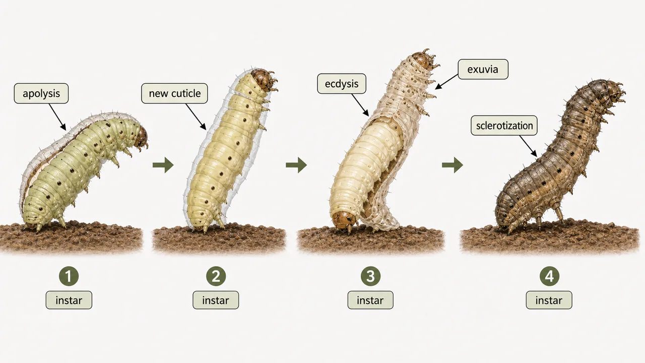
| Stadium | Time interval between two successive moults |
| Instar | The form of the insect during any stadium (e.g., 2nd instar = larva between 1st and 2nd moult) |
Three Stages of Moulting
Snake analogy: Moulting in insects works like a snake shedding skin -- but more complex. The insect builds a completely new suit of armour underneath the old one, then dissolves and recycles most of the old cuticle (saving nutrients), and finally breaks free. The whole process has three stages:
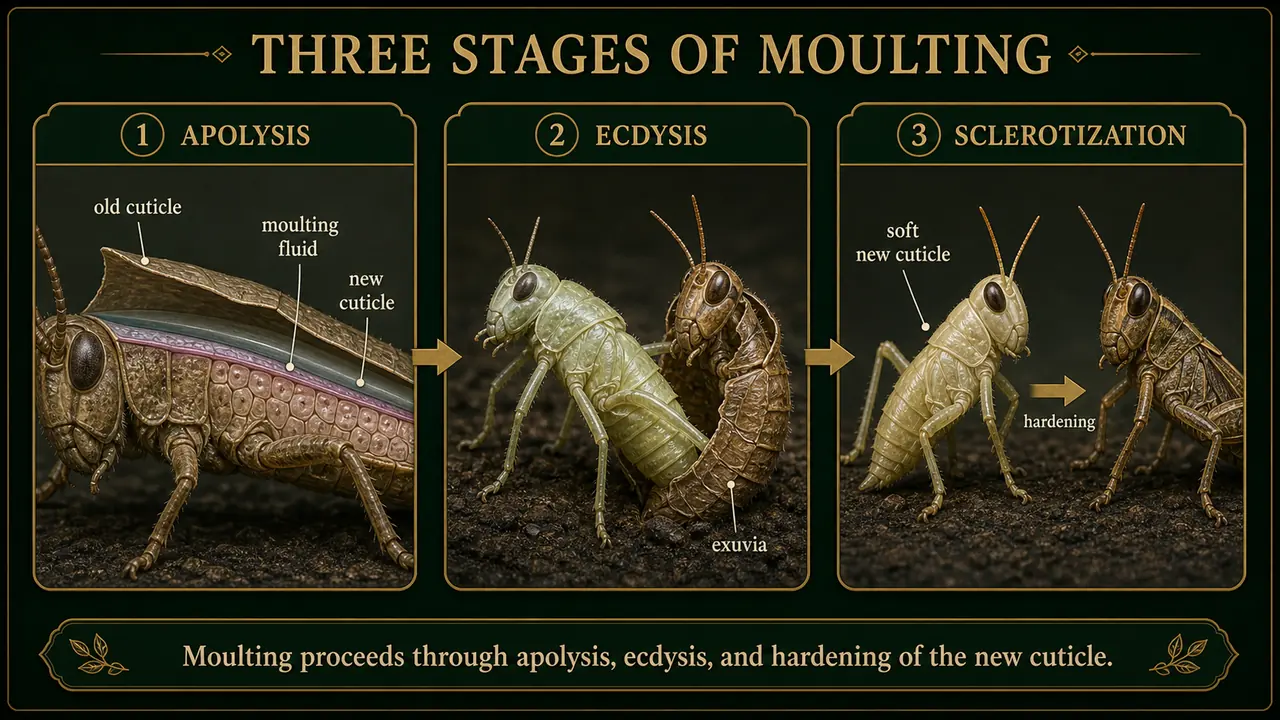
1. Apolysis (dissolution of old cuticle + formation of new)
- Epidermal cells undergo mitotic division, becoming columnar and closely packed.
- Increased cell size creates tension, separating epidermis from old cuticle.
- A sub-cuticular space forms; epidermal cells secrete moulting fluid containing two enzymes:
- Proteinase -- digests protein
- Chitinase -- digests chitin
- In older exam phrasing, this enzyme-rich secretion may also be called the moulting gel.
- These enzymes dissolve the endocuticle (recycling nutrients) while the new epicuticle and procuticle are deposited beneath.
- Undigested old exocuticle and epicuticle remain as the ecdysial membrane.
2. Ecdysis (shedding of old cuticle)
- The insect with both new cuticle underneath and old cuticle on top is called a pharate instar ("wearing two suits").
- The ecdysial membrane splits along the line of weakness (epicranial suture) due to:
- Muscular activity
- Swallowing air/water (distending the gut)
- Pumping blood from abdomen to thorax
- The new instar emerges -- head first, then thorax, abdomen, and appendages.
- The shed old cuticle is the exuviae.
3. Sclerotization (hardening of new cuticle)
- The soft, milky-white new cuticle becomes dark and hard through tanning (cross-linking of proteins with quinones) -- similar to how leather is tanned to become tough and durable.
- Procuticle differentiates into hard exocuticle and soft endocuticle.
- The insect is most vulnerable during this period -- soft body, pale colour, exposed to predators.
What a farmer sees in the field: A freshly moulted caterpillar appears pale, whitish, and sluggish for a few hours. Its body is visibly softer -- you can gently press it and feel the difference. This is the best window for contact sprays, as the protective wax layer is not yet fully formed. Within 4--6 hours, the cuticle darkens and hardens, and the insect becomes resistant again.
Agricultural relevance: Insect growth regulators (IGRs) like diflubenzuron target chitin synthesis during moulting, preventing the insect from forming a proper new cuticle. This is why IGRs are most effective against immature stages.
Moulting Hormones
Three hormones control moulting in a coordinated manner:
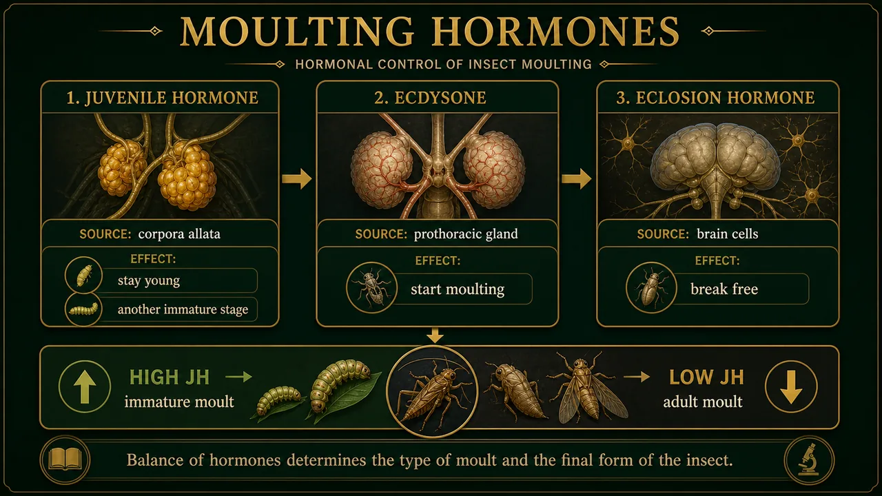
| Hormone | Produced By | Function | Memory Aid |
|---|---|---|---|
| Juvenile Hormone (JH) | Corpora allata (brain) | Keeps insect in immature stage; high JH = moult to another immature stage; low JH = moult to adult | "Stay young" signal -- like a parent saying "you're not old enough yet" |
| Moulting Hormone (Ecdysone) | Prothoracic glands | Triggers the moulting process by stimulating epidermal cells | "Start moulting" signal -- the alarm clock that wakes up the process |
| Eclosion Hormone | Neurosecretory cells (brain) | Triggers behaviours for breaking free from old cuticle (air swallowing, muscular contractions) | "Break free" signal -- the final push to escape the old shell |
How the three hormones work together: Imagine a growing child in a school uniform. JH decides "still a child, keep wearing uniform" (immature stage). Ecdysone says "time to get a new, bigger uniform" (triggers moulting). When JH finally drops low enough, ecdysone triggers the final moult into adult clothes (metamorphosis). Eclosion hormone is the burst of energy to actually change clothes.
Exam tip: JH from corpora allata (think "Allata = Adolescence"). Ecdysone from prothoracic glands (think "Prothoracic = Promotes moulting").
Summary Cheat Sheet
| Concept | Key Detail |
|---|---|
| Integument layers (outside in) | Cuticle → Epidermis (only living layer) → Basement membrane |
| Cuticle composition | Chitin (35--60%) + Protein (25--40%) |
| Epicuticle layers | Cement → Wax → Polyphenol → Cuticulin |
| Wax layer function | Waterproofing (prevents desiccation) |
| Procuticle parts | Exocuticle (hard, not digested in moulting) + Endocuticle (soft, recycled) |
| Key proteins | Arthropodin (soft), Sclerotin (hard), Resilin (elastic) |
| Chitin | Nitrogenous polysaccharide; 2nd most abundant after cellulose |
| Setae produced by | Trichogen cell (hair) + Tormogen cell (socket) |
| Chaetotaxy | Study of setal arrangement; useful for larval identification |
| Apodemes vs. Apophyses | Hollow (muscle attachment) vs. Solid (organ support) |
| Moulting stages | Apolysis → Ecdysis → Sclerotization |
| Exuvia / Stadium / Instar | Shed cuticle / Time between moults / Form during a stadium |
| Moulting enzymes | Proteinase + Chitinase (in moulting fluid) |
| JH source | Corpora allata |
| Ecdysone source | Prothoracic glands |
TIP
Next: The next lesson examines the insect head -- its sclerites, sutures, orientation types, and antenna modifications -- the sensory command centre of every insect.
References
1 source
References
Lesson Doubts
Ask questions, get expert answers