👹 Insect Head -- Structure, Orientation, and Antennae
Head capsule sclerites and sutures, three types of head orientation (hypognathous, prognathous, opisthognathous), antenna structure and modifications with agricultural examples
Chordoton organ found in Male Mosquito?
In the previous lesson, we covered the insect exoskeleton and moulting -- the body wall that protects insects and the process by which they grow. Now we move to the first body region: the head, the sensory and feeding command centre.
When a cotton farmer notices tiny green insects clustered on leaf undersides and wonders how they feed, the answer begins with the insect's head. The head houses the mouthparts that determine whether a pest chews (like a bollworm caterpillar) or sucks (like an aphid), and the antennae that guide it to the crop in the first place. Understanding head morphology is therefore the foundation of pest identification.
This lesson covers:
- Head structure -- seven fused segments, sclerites, and sutures
- Head orientation -- hypognathous, prognathous, and opisthognathous types
- Antennae -- structure, 14 modifications, and their functions
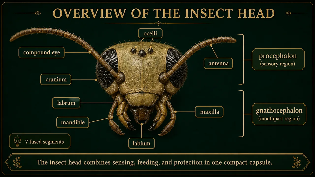
Overview of the Insect Head
Think of it this way: The insect head is like a helmet with built-in tools -- it has cameras (eyes), radar dishes (antennae), and power tools (mouthparts) all fused into one compact unit. Unlike our skull (which only protects the brain), the insect head is both a sensory hub and a feeding machine.
- The insect body is divided into three tagmata: head, thorax, and abdomen.
- This grouping of segments is called tagmosis.
- The head is a hard, highly sclerotized (hardened through protein cross-linking) compact structure.
- It consists of compound eyes, simple eyes (ocelli), mouthparts, and a pair of antennae.
- The head is the sensory and feeding centre of the insect.
- Formed by the fusion of 7 segments into a head capsule (individual segment boundaries are no longer visible in adults -- like 7 rooms merged into one open hall).
- The head is attached to the thorax by the cervix or neck region.
- Some older entomologists treat the adult head as effectively 6 visible segments because one segment becomes highly reduced during evolutionary fusion, but the standard teaching recall remains 7 segments.
- Head segments are divided into:
- Procephalon -- anterior segments bearing eyes and antennae (the "seeing and sensing" half)
- Gnathocephalon -- posterior segments bearing mouthparts (the "eating" half)
| S.N. | Segment | Head | Appendages |
|---|---|---|---|
| 1. | Pre Antennal Segment / Ocular | Procephalon | No appendages |
| 2. | Antennal Segment | Procephalon | Antennae |
| 3. | Intercalary Segment | Procephalon | No appendages |
| 4. | Clypeolabral Segment | Gnathocephalon | Labrum |
| 5. | Mandibular Segment | Gnathocephalon | Mandibles |
| 6. | Maxillary Segment | Gnathocephalon | Maxillae |
| 7. | Labial Segment | Gnathocephalon | Labium (lower lip) |
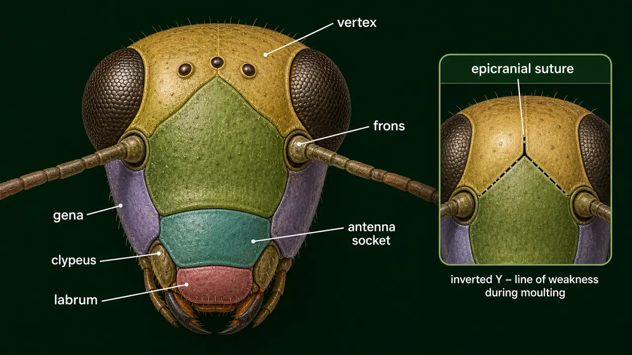
Head Sclerites (Regions of the Head Capsule)
The head capsule (excluding appendages) formed by fused sclerites is called the cranium. These sclerites are joined by cuticular ridges called sutures that provide mechanical support and muscle attachment.
- Sclerites are the hardened plates of the head capsule, while sutures are the softer lines or joints between adjacent sclerites.
| Sclerite | Location | Description |
|---|---|---|
| Vertex | Top of head | "Crown" of the head, between the two compound eyes |
| Frons | Facial region | "Forehead"; bears the median ocellus; important diagnostic feature |
| Clypeus | Above labrum | Divided into ante-clypeus (anterior) and post-clypeus (posterior); attachment point for labrum |
| Labrum | Below clypeus | Small sclerite forming the upper lip; freely suspended from clypeus margin |
| Gena | Below compound eyes | "Cheek" region, extending to just above the mandibles |
| Occular sclerites | Around compound eyes | Cuticular rings providing structural support to compound eyes |
| Antennal sclerites | Around antennal base (scape) | Well developed in Plecoptera (stone flies) |
| Sclerite | Location | Description |
|---|---|---|
| Epicranium | Upper head | Extends from vertex to occipital suture; covers dorsal and lateral surfaces |
| Occiput | Behind head | Inverted "U" shaped area between epicranium and post-occiput |
| Post-occiput | Extreme posterior | Rim around the occipital foramen (the opening through which aorta, foregut, nerve cord, and neck muscles pass) |
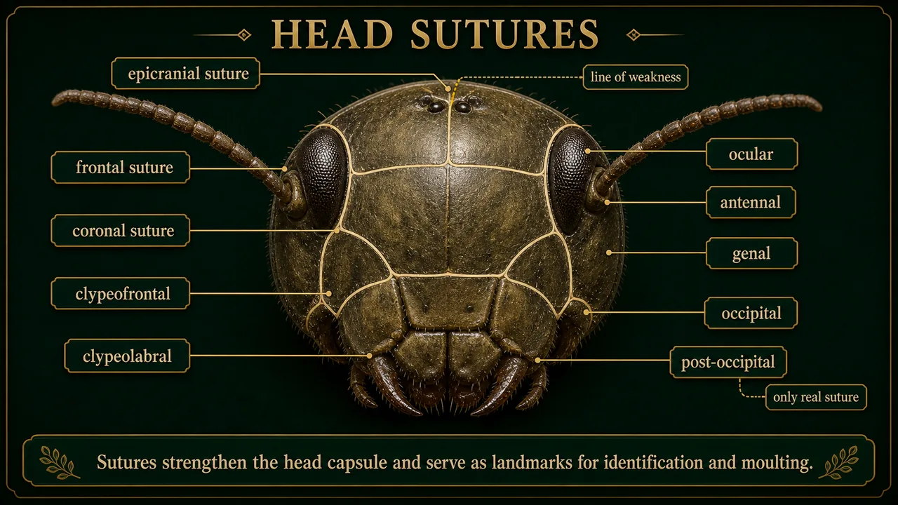
Head Sutures
Sutures (Latin: "joint") are cuticular lines that provide muscle attachment and serve as landmarks for identification.
| Suture | Shape | Location | Key Significance |
|---|---|---|---|
| Epicranial suture | Inverted Y | Above facial region to epicranium | Also called ecdysial suture or line of weakness -- the old cuticle splits here during moulting |
| Clypeolabral suture | -- | Between clypeus and labrum | Allows labrum to move freely for food manipulation |
| Clypeofrontal (epistomal) suture | -- | Between clypeus and frons | Important landmark for distinguishing facial regions |
| Occipital suture | U-shaped / horseshoe | Between epicranium and occiput | Marks rear boundary of main head capsule |
| Post-occipital suture | -- | Posterior end of head | The only real suture (represents true boundary between original segments); separates head from neck |
| Genal suture | -- | Lateral side (gena) | On the "cheek" region |
| Occular suture | Circular | Around each compound eye | -- |
| Antennal suture | Ring-shaped | Around antennal socket | Allows antenna to rotate and move in multiple directions |
Exam favourite: The epicranial suture is an inverted Y consisting of two frontal suture arms and a coronal suture stem. It is the line of weakness where cuticle splits during ecdysis.
Internal Structures and Functions
- Occipital foramen: Posterior opening through which aorta, foregut, central nerve cord, and neck muscles pass -- the vital gateway connecting head to body.
- Tentorium: Internal skeletal framework made of cuticular invaginations; acts as a brace inside the head, supporting muscles for antennae and mouthparts, and protecting the brain.
- Cephalic appendages: Compound eyes, 0--3 ocelli, antennae, and mouthparts.
- The maximum number of ocelli in an adult insect is three.
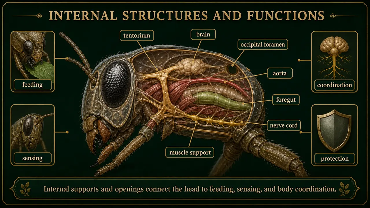
Four Functions of the Head
- Food ingestion -- mouthparts acquire and process food
- Sensory perception -- eyes, ocelli, antennae detect light, chemicals, sound, touch
- Coordination -- brain processes sensory information and coordinates responses
- Protection -- hard sclerotized capsule shields the brain and nerve centres
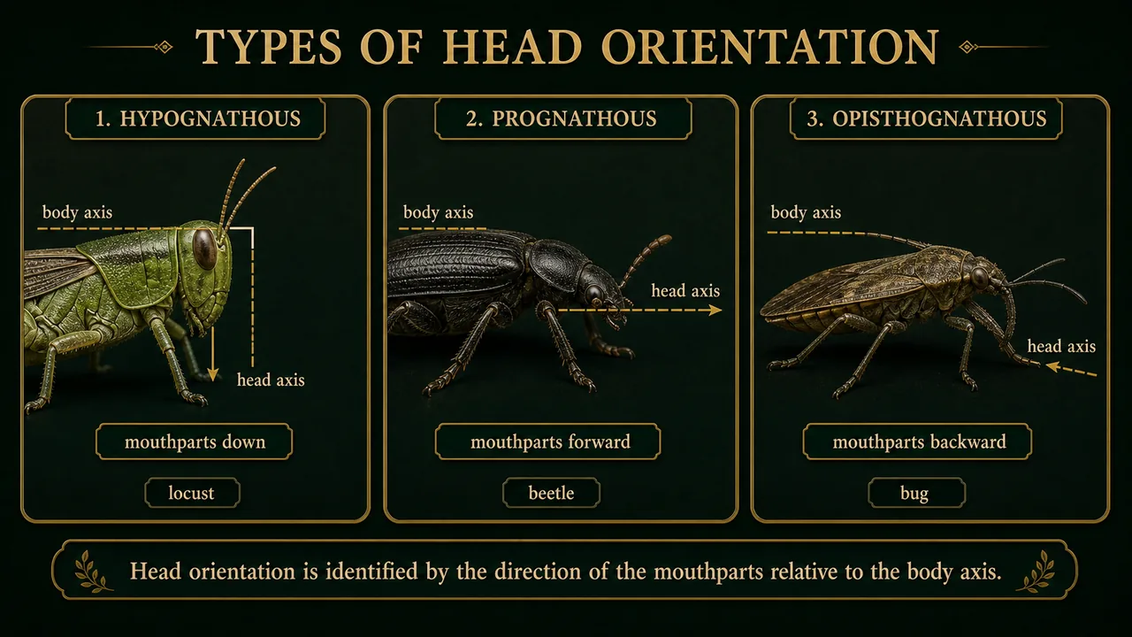
Types of Head Orientation
The position of the head relative to the body determines the direction of the mouthparts, reflecting the insect's feeding habits. This is a powerful diagnostic tool -- you can predict how an insect feeds just by looking at which way its head points.
| Type | Meaning | Mouthpart Direction | Body Angle | Also Called | Example | Agricultural Significance |
|---|---|---|---|---|---|---|
| Hypognathous | Hypo (below) + Gnathous (jaw) | Downward (ventral) | Head at right angle to body | Orthopteroid type | Locust, Grasshopper, Cockroach | Leaf-feeding pests that sit on top of leaves |
| Prognathous | Pro (forward) + Gnathous (jaw) | Forward | Head in same axis as body | Coleopteroid type | Beetles | Borers that tunnel into stems, wood, or soil |
| Opisthognathous | Opistho (behind) + Gnathous (jaw) | Backward (between forelegs) | Same as prognathous but directed backward | Hemipteroid / Opisthorhynchous | Bugs | Sap-sucking pests that insert stylets into plant tissue |
Extended head: Insects with a beak-like elongated head, such as Pyrilla perpusilla (sugarcane leafhopper), are termed opisthorhynchous -- the rostrum projects backward for phloem-feeding.
Reading shortcut: ignore the antennae first and look only at the mouthpart direction relative to the thorax. That single visual cue usually tells you the head orientation immediately.
Mnemonic: "H-P-O" = Hypognathous (down), Prognathous (forward), Opisthognathous (backward). The primitive condition is hypognathous.
Antennae
Antennae are among the most versatile sensory organs in the insect world -- they are the insect's nose, ears, and fingertips rolled into one. Knowing antenna type is a valuable tool for pest identification -- you can often identify an insect family just from its antenna shape.
- Single pair present in most insects.
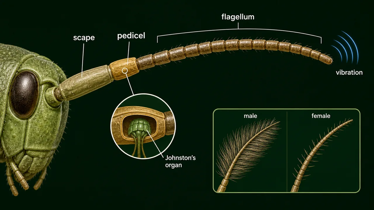
- Consist of three parts: scape (base), pedicel (middle), and flagellum (distal, many-segmented).
- Arise from the 2nd (antennal) segment of the head; nerves come from the deutocerebrum of the brain.
- Well developed in adults, poorly developed in immature stages.
- Base connected to the socket by an articulatory membrane allowing free movement.
Antenna Structure
| Part | Position | Key Feature |
|---|---|---|
| Scape | 1st (basal) segment | Largest and strongest; articulates with head via antennifer; moves the whole antenna |
| Pedicel | 2nd (middle) segment | Contains Johnston's organ -- a chordotonal (auditory) organ that detects vibrations and movement of flagellum; important for hearing and balance |
| Flagellum | 3rd (distal) segment | Most variable part; many sub-segments; where most sensory receptors (sensilla) are concentrated; bears chemoreceptors |
TIP
S-P-F (Scape-Pedicel-Flagellum) — think "SPF sunscreen" to recall order from base to tip. Note: Pedicel contains Johnston's organ (balance/hearing); Flagellum bears chemoreceptors.
Johnston's organ was discovered by American physician Christopher Johnston. In male mosquitoes, it is finely tuned to detect the wing-beat frequency of females for mate location.
WARNING
Order Protura — the only insects with no antennae. Their front legs possess many sensilla and function like antennae as sensory substitutes.
Modifications of Antennae
Antenna shape is closely related to the insect's lifestyle and sensory needs, making it a valuable identification tool.
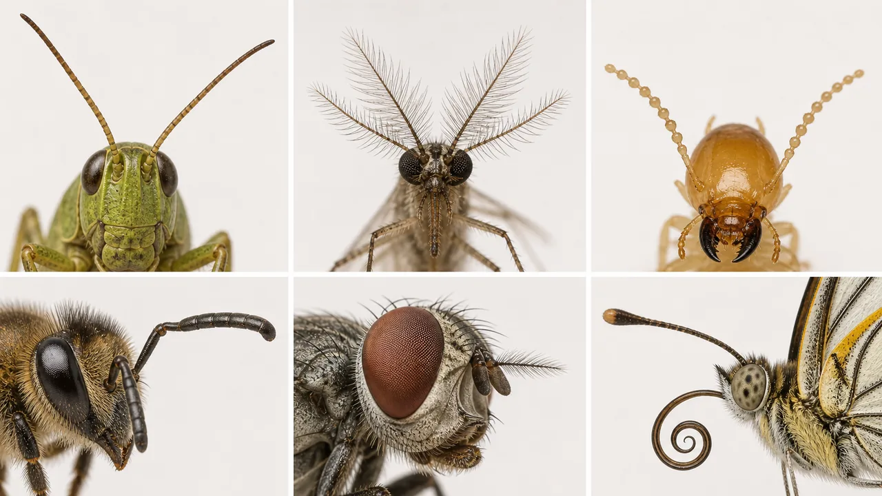
How to classify quickly: check whether the antenna is uniform, feathery, bead-like, elbowed, arista-bearing, or clubbed at the tip. Those shape cues are more reliable than trying to remember insect names first.
| Modification | Explanation | Examples |
|---|---|---|
| Filiform (Thread like) | Each segment is nearly equal in size and tapering to a point. Basically cylindrical in shape. | Grasshopper, Cockroach |
| Plumose (Feathery / Bushy) | Each segment bears a dense whorl of long, fine hair-like branches. Brush like structure. | Male mosquito |
| Moniliform (Bead like / Necklace like) | Each segment is round and roughly equal in thickness throughout, resembling a string of beads. | White ant (Termite) |
| Serrate (Saw like) | Each segment of antenna has a sharp, tooth like, triangular projecting process on one side. | Click beetle, Jewel beetle |
| Pectinate (Comb like) | Each segment has a long, slender lateral projection on one side, resembling the teeth of a comb. | Sawfly |
| Bipectinate (Double comb) | Each segment projects comb-like extensions on both sides, giving feathery appearance. | Male silkworm moth |
| Setaceous (Bristle like) | Segments gradually decrease in diameter from base to tip, tapering to a fine point (bristle-like). | Dragonfly, Grasshopper, Cricket |
| Pilose (Brush like with sparse hairs) | Looks like a plumose but each whorl contains a smaller number of hairs. | Female mosquito |
| Whorled | The whorl of bristles arises at every joint of segments. | Mango mealy bug |
| Clavate (Clubbed) | Segments gradually increase in diameter from base to tip ending in a club like apical part. | Khapra Beetle |
| Clavate with hook (Clubbed antennae with hook) | Segments gradually increase in diameter from base to tip and the last one ends with a small hook like structure. | Skipper butterflies |
| Capitate (Knobbed) | Terminal segments become enlarged suddenly and the terminal 3-5 segments suddenly enlarge to form a knob like structure. | Butterfly |
| Geniculate (Elbowed) | The first segment (scape) is long and apex form with small segments, hinged or bent like an elbow. | Honeybee, Ants, Weevils |
| Aristate (Antennae with arista) | Antennae are small, microscopic 3 segmented. 3rd segment enlarged and bears a bristle called arista on its dorsal side. | Housefly (Order: Diptera) |
| Lamellate (Plate like) | The terminal segments expand to one side and form broad plate or leaf like structure. | Rhinoceros beetles, Dung rollers, Chaffer beetles |
| Flabellate (Fan like) | Projections of some upper segments become long and form a feather like structure called flabella. | Stylopids |
| Stylate (Antennae with style) | Antennae small 3-4 segmented. Terminal segments elongate into a bristle like structure called style. | Robber fly |
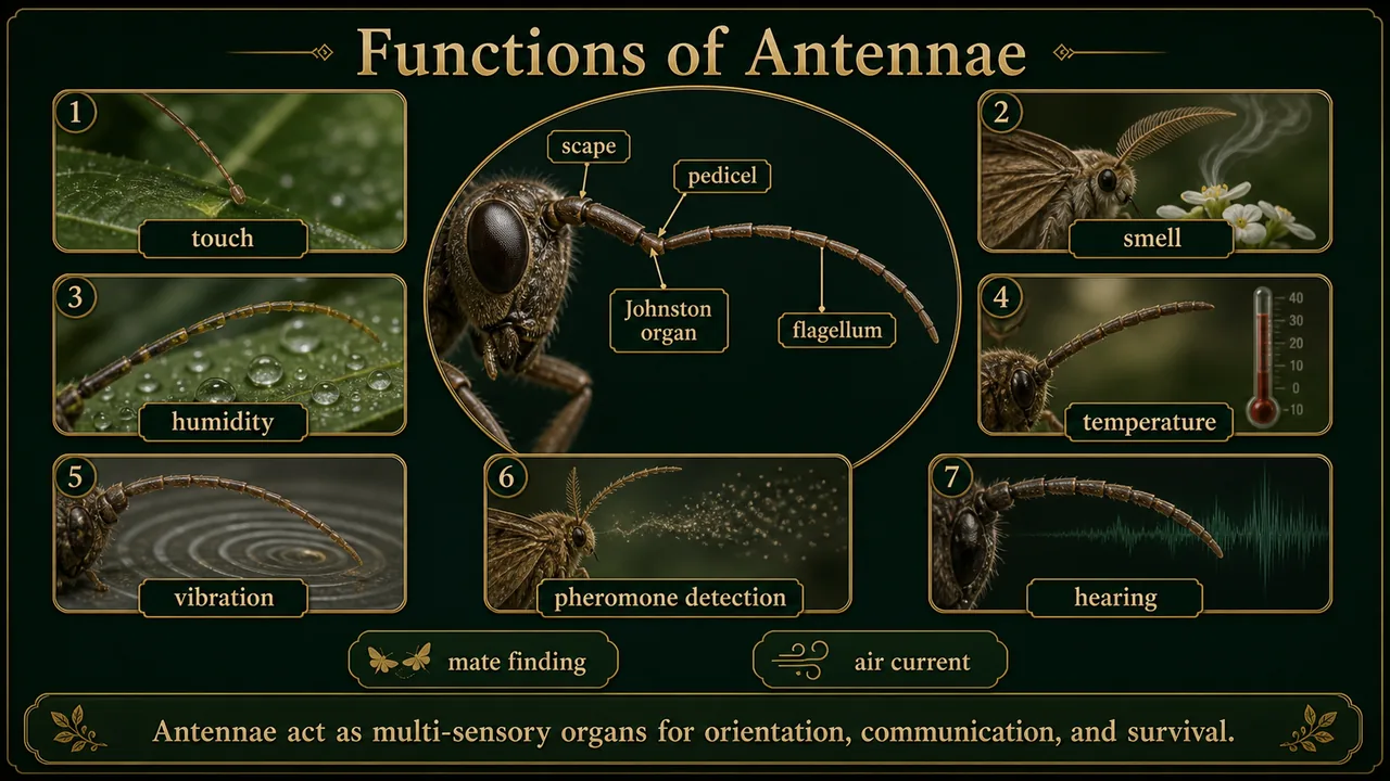
Functions of Antennae
| # | Function | Detail |
|---|---|---|
| 1 | Sensory organ | Responds to touch, smell, odour, humidity, temperature, vibration, wind velocity and direction |
| 2 | Chemical detection | Detects food and pheromones; male silk moths can find females up to 4.5 km away via sex pheromone -- large plumose antennae provide enormous surface area for chemosensory sensilla |
| 3 | Forward environment perception | Detects danger ahead |
| 4 | Hearing (Johnston's organ) | Auditory organ on pedicel; measures air current speed; in male mosquitoes, detects female wing-beat frequency |
| 5 | Sound production | Found in some insects (e.g., mole crickets) on the second segment |
| 6 | Prey handling | Helps mandibles hold prey and masticate food |
| 7 | Sexual dimorphism | Males and females often have different antenna shapes; males frequently have more elaborate antennae for pheromone detection |
| 8 | Clasping during copulation | Helps hold the female |
| 9 | Respiration aid | In aquatic insects, forms an air funnel channelling air from water surface to body |
Exam Tips
Epicranial suture shape: Inverted Y = coronal stem + two frontal arms. It is the line of weakness for ecdysis.
Post-occipital suture: The only real suture in the insect head -- separates head from neck and represents a true segmental boundary.
Johnston's organ location: Always on the pedicel (2nd antennal segment). Found in male mosquitoes (frequently tested).
Head orientation mnemonic: "Down-Forward-Back" = Hypognathous-Prognathous-Opisthognathous.
Summary Cheat Sheet
| Concept | Key Detail |
|---|---|
| Head segments | 7 fused segments forming cranium |
| Procephalon vs. Gnathocephalon | Eyes + antennae vs. mouthparts |
| Epicranial suture | Inverted Y; line of weakness for ecdysis |
| Only real suture | Post-occipital suture |
| Occipital foramen | Posterior opening connecting head to body |
| Tentorium | Internal skeleton for muscle attachment |
| Hypognathous | Mouthparts downward; primitive; e.g., grasshopper |
| Prognathous | Mouthparts forward; borers/predators; e.g., beetles |
| Opisthognathous | Mouthparts backward; sap-suckers; e.g., bugs |
| Antenna parts | Scape → Pedicel → Flagellum |
| Johnston's organ | On pedicel; hearing/balance; prominent in male mosquitoes |
| Deutocerebrum | Brain region processing antennal sensory information |
TIP
Next: The next lesson examines biting and chewing mouthparts -- the primitive mandibulate type found in grasshoppers, cockroaches, and caterpillars.
References
1 source
References
Lesson Doubts
Ask questions, get expert answers