🍄 Fungi, Algae, Viruses & Mycoplasma — Structure, Features, and Agricultural Significance
Key features, comparisons, and exam-critical facts about fungi, algae, viruses, mycoplasma, and their roles in agriculture with mnemonics and summary tables
From Field to Lab — Four Groups That Shape Every Farm
Picture a wheat field in Punjab after the monsoon. The leaves are covered with orange-brown pustules — that is rust, caused by a fungus. In a nearby rice paddy, the standing water has a greenish sheen — that is blue-green algae, a natural biofertiliser fixing nitrogen for free. A tomato plant in the kitchen garden has curled, yellowing leaves — the culprit is leaf curl virus, transmitted by whiteflies. And in the neighbouring brinjal field, plants with tiny, bunched leaves signal little leaf disease, caused by mycoplasma (phytoplasma).
These four groups — fungi, algae, viruses, and mycoplasma — together account for the vast majority of crop diseases and biological processes in agriculture. Understanding their differences is essential for both farm management and competitive exams.
Fungi — The Largest Group of Plant Pathogens
Fungi are eukaryotic, heterotrophic organisms. They cannot make their own food and depend on organic matter — either as saprophytes (feeding on dead matter) or as parasites (feeding on living hosts).
Structure and Key Features
| Feature | Detail |
|---|---|
| Cell type | Eukaryotic |
| Nutrition | Heterotrophic (cannot photosynthesize) |
| Size | 1.5–10 microns |
| Single filament | Hypha (plural: hyphae) |
| Group of hyphae | Mycelium |
| Root-like structures of Rhizopus | Rhizoids |
| Nutrient-absorbing structures in host | Haustoria |
| Best culture media | Potato Dextrose Agar (PDA) |
| Solidifying agent in media | Agar-agar |
| Yeast reproduction (asexual) | Budding |
| Yeasts are responsible for | Fermentation |
Additional Fungal Facts Asked in Exams
- A single fungal filament is a hypha, while the full fungal colony is the mycelium.
- The fungal classes grouped as Deuteromycetes are often called imperfect fungi because sexual reproduction is absent from the observed life cycle.
- In Oomycetes, the cell wall is rich in cellulose rather than chitin.
- Important asexual fungal fruiting bodies include pycnidia and acervuli.
Nutrition-Based Classification of Fungi
| Type | Meaning | Examples |
|---|---|---|
| Obligate saprophytes | Strictly depend on dead or decaying organic matter | Many decomposer moulds such as Rhizopus |
| Facultative saprophytes | Primarily parasitic forms that can continue growth saprophytically on dead tissue or culture media | Some plant-pathogenic fungi after host tissue dies |
| Obligate parasites | Survive only on living hosts | Puccinia |
| Facultative parasites | Mostly saprophytic but can parasitize | Fusarium |
| Symbiotic fungi | Live in beneficial association | Mycorrhiza |
- A good way to remember the two facultative categories is to ask what the fungus is mostly adapted to: a facultative parasite is mainly saprophytic but can attack a host, whereas a facultative saprophyte is mainly parasitic but can continue on dead organic matter.
Simplified Fungal Groups to Remember
| Group | Sexual spores | Typical note |
|---|---|---|
| Zygomycetes | Zygospores | Bread mould type group; asexual reproduction commonly occurs by sporangiospores |
| Ascomycetes | Ascospores | Sac fungi |
| Basidiomycetes | Basidiospores | Smuts, rusts, mushrooms |
| Deuteromycetes | Sexual stage absent | Imperfect fungi; asexual reproduction commonly by conidia / conidiospores |
| Oomycetes | Oospores | Cellulose-rich wall |
- In basic fungal reproduction language, sexual reproduction happens by the fusion of compatible sex organs or gametes, and the terminology depends on whether the two participating cells look alike or different.
- When morphologically similar gametes fuse, the process is described as isogamy. When a larger non-motile female structure and a smaller male structure are involved, the process is described as oogamy, and it ultimately leads to formation of an oospore.
- In some lower fungal groups, sexual reproduction may also be described through the fusion of two gametangia, helping students connect the reproductive process with the sexual spore that is finally produced.
- In exam-style fungal classification, it helps to pair each group with its usual spore cue: zygomycetes with sporangiospores in asexual reproduction, deuteromycetes with conidia / conidiospores, and the sexual spores as the classical group-defining markers.
Common Microbial Toxin Recalls
- Tentoxin is classically linked with Alternaria alternata.
- Fusaric acid is a standard toxin recall for Fusarium species.
- Aflatoxins are linked with Aspergillus flavus and related species.
IMPORTANT
All fungi are heterotrophs while all algae are autotrophs. This is the single most important distinction between these two groups for exams.
TIP
Mnemonic — "Fungi Hunt, Algae Auto": Fungi are Heterotrophs (they hunt for food), Algae are Autotrophs (they auto-produce food via photosynthesis).

Algae — The Photosynthetic Microorganisms
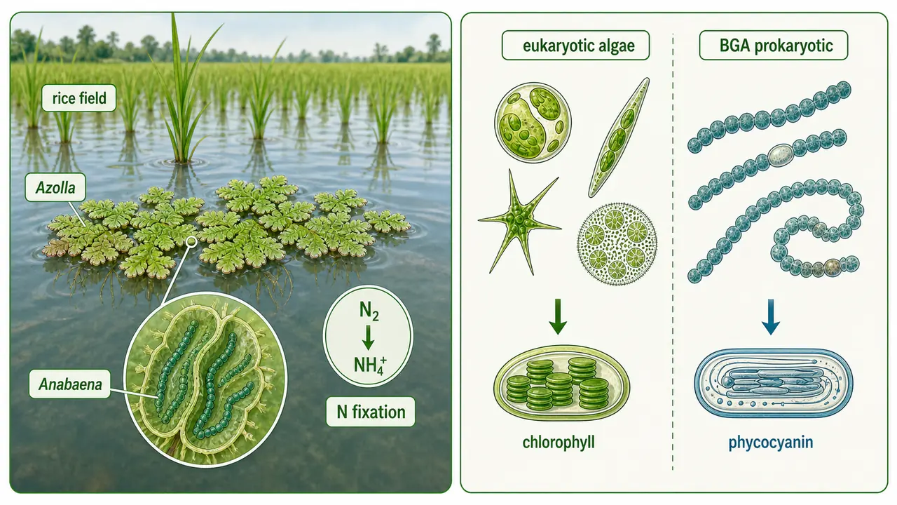
Algae are photosynthetic microorganisms that are nutritionally Autotrophs. They are also called primary producers of organic matter because they convert sunlight into food.
Key Features
| Feature | Detail |
|---|---|
| Nutrition | Autotrophic (photosynthetic) |
| Growth requirement | Can grow only in Light |
| Cell type | Eukaryotic (except BGA which are prokaryotic) |
Blue-Green Algae (BGA) — A Special Case
BGA (Cyanobacteria) are unique because they are prokaryotic organisms that contain Chlorophyll — making them the only prokaryotes capable of oxygenic photosynthesis.
| Feature | Detail |
|---|---|
| Cell type | Prokaryotic (unlike other algae) |
| Blue pigment | Phycocyanin |
| Agricultural use | Biofertiliser in rice fields |
| N-fixation association | Anabaena (BGA) + Azolla (water fern) |
TIP
BGA in rice: The Anabaena-Azolla association fixes nitrogen in flooded rice fields. Anabaena is the BGA that lives inside the water fern Azolla. This symbiosis can fix 20–40 kg N/ha/season — a frequently asked fact.
- In compact algae-classification recall, green algae are linked with Chlorophyta, golden-brown algae / diatoms with Bacillariophyta, and yellow-green algae with Xanthophyta.
Viruses — The Smallest Infectious Agents
Viruses are smaller than bacteria and are obligate intracellular parasites — they can only multiply inside living host cells. Outside a host, they exist as inert chemical particles.
Key Features
| Feature | Detail |
|---|---|
| Size | 0.06–0.14 microns (smallest of all microorganisms) |
| Parasitism | Obligate intracellular (must be inside a living cell) |
| Crystalline nature proved by | Stanley |
| Viruses that infect bacteria | Bacteriophages |
Classic Virus History Recalls
- Adolf Mayer first described tobacco mosaic disease in 1882 and demonstrated its sap transmissibility.
- Ivanowsky (1892) showed that the infectious agent causing tobacco mosaic disease was filterable.
- M.W. Beijerinck later treated it as a new type of infectious entity and popularised the classical virus idea as a contagious living fluid. In direct history recall, Beijerinck (1898) is also linked with the term virus.
- In standard exam one-liners, M.W. Beijerinck is also remembered as the Father of Virology.
Virus Composition — A Critical Exam Table
Different types of viruses have different chemical compositions. This is one of the most frequently tested topics.
- The protein coat of a virus is called the capsid.
- The viral nucleic acid together with its capsid is referred to as the nucleocapsid.
- A complete mature infectious virus particle is called a virion.
| Type | Composition | Memory Aid |
|---|---|---|
| Plant virus | RNA + Protein | Plant = RNA (both have "a" sound) |
| Animal virus | DNA + Protein | Animal = DNA |
| Lipo-virus | Nucleic acid + Protein + Lipid | Has a lipid envelope |
| Viroid | RNA only (naked nucleic acid, no protein coat) | Viroid = "void" of protein |
| Virusoid | Small satellite-like RNA associated with a helper virus | Even simpler subviral RNA partner |
| Prion | Proteinaceous infectious particle with no nucleic acid | Even simpler than viruses in structure |
IMPORTANT
Plant viruses mostly contain RNA, while animal viruses (and bacteriophages) contain DNA. Viroids are even simpler — just naked RNA without any protein coat. Virusoids are small subviral RNAs that depend on a helper virus, while prions are infectious proteins without nucleic acid. Viroids cause diseases like potato spindle tuber and coconut cadang-cadang.
- In standard plant-pathology recall, viroid diseases are classically described as being transmitted mainly by sap contact or mechanical means, while specific insect vectors are generally not emphasized for them.
- In direct history recall, prions are linked with Stanley Prusiner (1982).
- Standard disease examples associated with prions include mad cow disease and scrapie of sheep and goats.
- In direct exam shorthand, prions are often treated as the smallest infectious agents, with viroids usually placed next after them in minimal-pathogen comparisons.
NOTE
Older exam notes also contrast smallest living infectious agent = mycoplasma against smallest infectious agent overall = prion, with viroids placed just after prions in the ultra-small non-cellular series.
- Bacteriophages are viruses that infect bacteria. They were first recognized through the work of Twort (1915) and d'Herelle (1917), and the term bacteriophage itself is classically associated with Felix d'Herelle.
- In compact bacteriophage classification, a lytic phage is the classically virulent type, while a temperate phage is the classically avirulent / lysogenic-capable type; older objective recall often pairs T-phage with the lytic type and lambda phage with the temperate type.
NOTE
In comparative exam lists, the usual order from larger classical microbes to the smallest non-cellular agents is often stated as: Algae → Protozoa → Fungi → Actinomycetes → Bacteria → Mycoplasma → Virus → Viroid → Prion.

Mycoplasma (MLO / Phytoplasma) — The Wall-less Pathogens
Mycoplasma (Mycoplasma-Like Organisms) are unique pathogens that are larger than viruses but smaller than bacteria. They cause many important yellowing diseases in plants.
In older microbiology and plant-pathology recall tables, this wall-less group is also linked with the term PPLO — Pleuro-Pneumonia-Like Organism — which was historically used for the mycoplasma group before the current plant-focused MLO / phytoplasma wording became more common.
In plant pathology, phytoplasmas and spiroplasmas are both recognized as phloem-inhabiting, wall-less mollicutes. They are grouped near mycoplasma because they share the same general biological logic of reduced structure and dependence on living host tissues.
Key Features
| Feature | Detail |
|---|---|
| Size | 0.1–0.3 microns |
| Cell wall | Devoid of cell wall (no cell wall) |
| Cell boundary | Flexible triple-layered cell membrane instead of a rigid cell wall |
| Shape | Highly Pleomorphic (variable shape) |
| Nucleic acid | Contains both DNA and RNA |
| Resistant to | Penicillin (targets cell wall — mycoplasma has none) |
| Sensitive to | Tetracycline (targets protein synthesis) |
- In standard microbiology recall, cultured mycoplasma colonies are often described as having a fried-egg appearance, reflecting their dense centre and spreading margin.
- In plant-disease transmission recall, many important phytoplasma diseases are classically associated with leafhopper vectors, matching their phloem-limited habit.
- In older objective plant-pathology history, mulberry dwarf is commonly cited as the first phytoplasma disease, linked with Doi and Ishii (1967).
Spiroplasma
- Spiroplasma are helical, wall-less mollicutes related to the mycoplasma group.
- Like phytoplasmas, they inhabit the phloem and depend on living host tissues for survival and spread.
- Two standard textbook examples are:
- Corn stunt as the first widely remembered spiroplasma disease
- Citrus stubborn as another important spiroplasma-associated disease
WARNING
Penicillin vs Tetracycline — The No. 1 Mycoplasma Exam Question:
- Mycoplasma has no cell wall
- Penicillin works by disrupting cell wall synthesis — so it is ineffective against mycoplasma
- Tetracycline inhibits protein synthesis — so it works against mycoplasma
- This is tested in nearly every agriculture competitive exam.
Important Mycoplasma Diseases
| Crop | Disease |
|---|---|
| Brinjal | Little leaf |
| Sesamum | Phyllody |
| Sandal | Spike disease |
| Sugarcane | Grassy shoot |
| Potato | Purple top / Witches' broom |
Comparison Table — Fungi vs Algae vs Viruses vs Mycoplasma
| Feature | Fungi | Algae | Viruses | Mycoplasma |
|---|---|---|---|---|
| Cell type | Eukaryotic | Eukaryotic (BGA: Prokaryotic) | Acellular | Prokaryotic |
| Nutrition | Heterotrophic | Autotrophic | Obligate parasite | Parasitic |
| Size | 1.5–10 microns | 0.1+ microns | 0.06–0.14 microns | 0.1–0.3 microns |
| Cell wall | Present (chitin) | Present (cellulose) | Absent | Absent |
| Nucleic acid | DNA + RNA | DNA + RNA | DNA or RNA | DNA + RNA |
| Reproduction | Spores, budding | Binary fission, spores | Inside host only | Binary fission |
| Culture on artificial media | Yes (PDA) | Yes | No (obligate parasite) | Difficult |
| Antibiotic sensitivity | Fungicides | — | No antibiotic works | Tetracycline |
Small but Important Soil-Microbiology Recalls
- Actinomycetes are filamentous bacteria often remembered as ray fungi in older agricultural microbiology notes.
- The characteristic earthy smell of freshly wetted soil is classically linked with actinomycetes, especially through the compound geosmin.
- Among soil protozoa, the classically emphasized dominant group in old exam recall is Mastigophora.
Oxidative vs Photophosphorylation
These two processes are the main ways living cells produce ATP (energy currency).
| Feature | Oxidative Phosphorylation | Photophosphorylation |
|---|---|---|
| Occurs in | Mitochondria | Chloroplasts |
| Energy source | Chemical energy (from food breakdown) | Light energy (from sun) |
| Process | Part of respiration | Part of photosynthesis |
| Common mechanism | Chemiosmotic coupling | Chemiosmotic coupling |
TIP
Both processes use chemiosmotic coupling (proton gradient across a membrane drives ATP synthase). The difference is the energy source: food (mitochondria) vs light (chloroplasts).
Summary Table — Key Facts at a Glance
| Fact | Answer |
|---|---|
| All fungi are | Heterotrophs |
| All algae are | Autotrophs |
| BGA cell type | Prokaryotic |
| Blue pigment in BGA | Phycocyanin |
| N-fixation in rice fields | Anabaena + Azolla |
| Smallest microorganism | Viruses (0.06–0.14 microns) |
| Plant virus composition | RNA + Protein |
| Animal virus composition | DNA + Protein |
| Viroid composition | RNA only |
| Crystalline nature of viruses | Stanley |
| Viruses infecting bacteria | Bacteriophages |
| Mycoplasma has no | Cell wall |
| Mycoplasma resistant to | Penicillin |
| Mycoplasma sensitive to | Tetracycline |
| Spiroplasma habitat | Phloem tissue |
| Classic spiroplasma diseases | Corn stunt, Citrus stubborn |
| Haustoria are | Nutrient-absorbing fungal structures in host |
| Best media for fungi | PDA (Potato Dextrose Agar) |
| Solidifying agent | Agar-agar |
| Yeast reproduction | Budding |
| Both phosphorylation types use | Chemiosmotic coupling |
Summary Cheat Sheet
| Fact | Answer |
|---|---|
| Fungi cell type | Eukaryotic |
| Fungi nutrition | Heterotrophic |
| Single fungal filament | Hypha |
| Group of hyphae | Mycelium |
| Root-like structures in Rhizopus | Rhizoids |
| Nutrient-absorbing fungal structures | Haustoria |
| Best media for fungi | PDA (Potato Dextrose Agar) |
| Solidifying agent in media | Agar-agar |
| Yeast reproduction | Budding |
| Imperfect fungi | Deuteromycetes |
| Oomycete wall material | Cellulose |
| All algae are | Autotrophs |
| BGA cell type | Prokaryotic |
| Blue pigment in BGA | Phycocyanin |
| N-fixation in rice fields | Anabaena (BGA) + Azolla (water fern) |
| Smallest microorganism | Viruses (0.06–0.14 microns) |
| Viruses are obligate | Intracellular parasites |
| Plant virus composition | RNA + Protein |
| Animal virus composition | DNA + Protein |
| Viroid composition | RNA only (no protein coat) |
| Prion composition | Protein only |
| Crystalline nature of viruses proved by | Stanley |
| Viruses infecting bacteria | Bacteriophages |
| Mycoplasma has no | Cell wall |
| Mycoplasma resistant to | Penicillin |
| Mycoplasma sensitive to | Tetracycline |
| Mycoplasma size | 0.1–0.3 microns |
| Spiroplasma | Helical phloem-inhabiting wall-less mollicutes |
| Little leaf disease (brinjal) caused by | Mycoplasma (Phytoplasma) |
| Both phosphorylation types use | Chemiosmotic coupling |
Lesson Doubts
Ask questions, get expert answers