🍽️ Insect Digestive System Structure
Structure and functions of the alimentary canal and digestive organs in insects.
The insect digestive system is built around a tubular alimentary canal that runs from mouth to anus. Its structure changes according to feeding habit, but the same broad plan is seen across most insects: foregut, midgut, and hindgut, along with digestive glands, enzymes, and microbial helpers.
Structure and Functions of the Digestive System
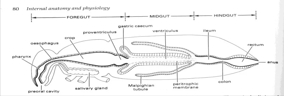
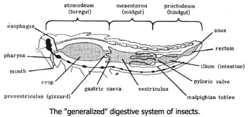
The alimentary canal is long, muscular, and tubular. It performs ingestion, storage, grinding, digestion, absorption, and finally elimination of wastes. The canal is divided into:
- Foregut
- Midgut
- Hindgut
Foregut
The foregut is ectodermal in origin and is also called the stomodeum. It has an internal cuticular lining. The terminal mouthparts open into the preoral cavity.
- The space between epipharynx and hypopharynx is the cibarium.
- The space between hypopharynx and the salivary duct is the salivarium.
- The pharynx is a muscular organ that pushes food into the oesophagus and acts as a sucking pump in sap feeders.
- The oesophagus is a narrow tube conducting food to the crop.
- The crop is the dilated distal part of the oesophagus and acts as a food reservoir.
- In bees, the crop functions as a honey stomach.
- The proventriculus or gizzard is the posterior part of the foregut and is well muscled.
The gizzard is found mainly in solid feeders and is reduced or absent in fluid feeders. Its cuticle may be modified into:
- Teeth, as in cockroach, for grinding and straining food
- Plates, as in honey bee, for separating pollen from nectar
- Spines, as in flea, for breaking blood corpuscles
Food passes from foregut to midgut through the cardiac valve or oesophageal valve.
Midgut
The midgut is endodermal in origin and is also called the mesenteron. It lacks cuticular lining and is the chief site of digestion and absorption.
The midgut epithelium includes:
- Secretory or columnar cells
- Goblet cells
- Regenerative cells, which replace worn-out secretory cells
Important structures of the midgut
1. Peritrophic membrane This is a semipermeable internal lining secreted by midgut epithelial cells. It is common in solid feeders and generally absent in sap feeders.
Functions:
- Lubricates and facilitates food movement
- Envelops food
- Protects midgut epithelium from hard food particles
2. Gastric caecae Also called enteric or hepatic caecae, these are finger-like outgrowths near the anterior or posterior region of the midgut. They increase digestive surface area and may shelter symbiotic microorganisms.
3. Pyloric valve Also called the proctodeal valve, it regulates food passage from midgut to hindgut. In some immature insects, the midgut and hindgut remain disconnected until pupation. Example: honey bee grub.
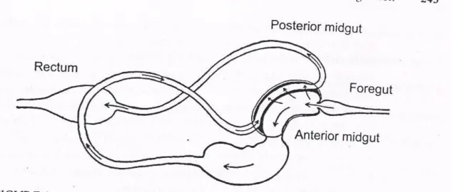
4. Filter chamber This is a specialized arrangement in which parts of the midgut and hindgut lie close together inside a common sac. It is common in Homopteran liquid feeders and helps:
- Remove excess water from dilute food
- Prevent dilution of digestive enzymes
- Concentrate nutrients
- Support osmoregulation
Hindgut
The hindgut is ectodermal in origin and develops from posterior invagination of the ectoderm. Like the foregut, it has a cuticular lining.
Its main functions are:
- Absorption of water
- Absorption of salts, ions, amino acids, and other useful substances
- Formation and elimination of faeces
The hindgut is divided into:
- Ileum
- Colon
- Rectum
In larvae of scarabids and termites, the ileum is pouch-like and acts as a fermentation chamber for symbionts. The rectum contains rectal pads, which help in dehydration of faeces before expulsion through the anus.
Gut Physiology
The main functions of the gut are digestion of ingested food and absorption of the resulting metabolites. Digestion is supported by enzymes, glands, and sometimes symbiotic microbes.
Digestive Glands
Salivary glands
In cockroach, a pair of labial glands functions as salivary glands and opens into the salivarium. In caterpillars, the mandibular glands secrete saliva, while the salivary glands are modified for silk production.
Functions of saliva
- Moistens and dissolves food
- Lubricates mouthparts
- Stimulates gustatory receptors
- In cockroach, contains amylase for starch digestion
- In honey bee, contains invertase for sucrose digestion
- In jassid, contains lipase and protease and may also contain toxins that cause tissue necrosis and phytotoxemia
- In plant bugs, contains pectinase that helps in stylet penetration and extra-intestinal digestion
- In mosquitoes, contains anticoagulant substances preventing blood clotting
- In gall midge, may contain indole acetic acid (IAA), which induces gall formation
- In vectors, saliva may help in pathogen transmission
Hepatic caecae and midgut epithelial cells
These structures secrete most digestive juices. Two modes of enzyme secretion are recognized:
- Holocrine secretion: the epithelial cell disintegrates during secretion
- Merocrine secretion: secretion occurs without cell breakdown
Digestive Enzymes
| Insect group | Enzyme | Substrate |
|---|---|---|
| Phytophagous larvae | Amylase | Starch |
| Maltase | Maltose | |
| Invertase | Sucrose | |
| Omnivorous insects | Protease | Protein |
| Lipase | Lipid | |
| Nectar feeders | Invertase | Sucrose |
| Wood-boring cerambycid grub and termites | Cellulase | Cellulose |
| Meat-eating maggots | Collagenase | Collagen and elastin |
| Bird lice | Keratinase | Keratin |
Microbes in Digestion
Some insect cells house symbiotic microorganisms. Such cells are called mycetocytes, and their aggregation forms a mycetome.
Important examples:
- Flagellate protozoa produce cellulase in termites and wood cockroach
- Bacteria help in wax digestion in wax moth
- Bed bugs and cockroaches obtain vitamins and amino acids from symbiotic microbes
Transmission may occur through:
- Trophallaxis: mouth-to-mouth food exchange
- Transovarial transmission: passage through the egg
In some insects such as plant bugs and antlion grubs, part of digestion occurs in the host or prey body before food is actually ingested. This is called extra-intestinal digestion.
Absorption
In most insects, nutrients are absorbed through the microvilli of midgut epithelial cells by diffusion. Water and ions are largely absorbed through the rectum.
Special cases include:
- Cockroach: lipid absorption occurs through the crop
- Termites and scarabaeid larvae: absorption may also occur through the ileum
- Solid feeders: water is resorbed in the rectum, and faeces are expelled as pellets
- Sap feeders: faeces remain liquid
The sugar-rich liquid excreta of homopteran bugs such as aphids, mealybugs, scales, and psyllids is called honeydew. It attracts ants and is a common field sign of infestation.
Alimentary System of Cockroach
Cockroach has a typical holotrophic alimentary canal extending from mouth to anus. It is divided into foregut, midgut, and hindgut.
- Mouth opens into pharynx
- Pharynx leads to oesophagus
- Oesophagus enlarges into crop for food storage
- Crop narrows into the gizzard or proventriculus
- The gizzard bears six chitinous teeth that help grind food
- Midgut is the chief region for digestion and assimilation
- Hindgut is divided into ileum, colon, and rectum
Associated digestive structures include:
- Salivary glands with reservoirs
- Hepatic caecae at the foregut-midgut junction
- Malpighian tubules at the midgut-hindgut junction, which remove nitrogenous wastes from the haemolymph
Summary Cheat Sheet
- Three main gut regions: Foregut, midgut, hindgut.
- Foregut: Ectodermal, cuticle-lined, includes pharynx, oesophagus, crop, and gizzard.
- Midgut: Endodermal, no cuticular lining, main site of digestion and absorption.
- Hindgut: Ectodermal, cuticle-lined, involved in water and salt absorption.
- Cibarium: Space between epipharynx and hypopharynx.
- Salivarium: Space associated with hypopharynx and salivary duct opening.
- Peritrophic membrane: Semipermeable midgut lining common in solid feeders.
- Filter chamber: Special adaptation in sap feeders for removing excess water.
- Rectal pads: Help dehydrate faeces.
- Mycetocyte/Mycetome: Symbiont-containing cell and organ.
- Honeydew: Sugar-rich liquid excreta of sap feeders.
- Exam tip: Learn the function of crop, gizzard, gastric caecae, filter chamber, and rectal pads with one insect example where relevant.
References
2 sources • [1] [2]
References
Fundamentals of Entomology
Insect Morphology and Systematics
Lesson Doubts
Ask questions, get expert answers