📈 Diseases of Bhendi
Diseases of Bhendi.
This lesson outlines major bhendi diseases and provides concise, exam-ready understanding of etiology, spread, and field management.
Bhendi
Cercospora Leaf Spots : Cercospora malayensis,
C. abelmoschi
Symptoms
In India, two species of Cercospora produce leaf spots in bhendi. C. Malayensis causes
brown, irregular spots and C . abelmoschi causes sooty black, angular spots.Both the leaf spots
cause severe defoliation and are common during humid seasons.
Pathogen
Conidiophores are pale to medium olivaceous brown, multiseptate, some times branched,
geniculate and irregular. Conidia are obclavate to cylindric, olivaceous brown and straight to
curved.
Mode of spread and survival
The fungus survives in the diseased crop material.
Management
Spraying Mancozeb 0.25 % control the disease.
Fusarium wilt : Fusarium oxysporum f.sp. vasinfectum
Symptoms
The conspicuous symptom is a typical wilt, beginning with a yellowing and stunting of
the plant, followed by wilting and rolling of the leaves as if the roots were unable to supply
sufficient water. Finally, the plant dies. If a diseased stem is split lengthwise, the vascular
bundles appear as dark streaks. When severely infected, nearly the whole stem is blackend.
Pathogen
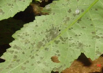 Macroconidia are 3- 5 septate formed on sporodochia and pionnotes. In mass conidia
appear buff or salmon orange in color. Macroconidia are fusiform and curved inward at both
ends. The base is pedicellate. Microconidia are septate. Terminal and intercalary chlamydospores
are broadly ovate.
Mode of spread and survival
The fungus is soil borne.
Management
Treat the seeds with Mancozeb @ 3g/kg seed. Drench the field with Copper oxy chloride
@ 0.25%.
Powdery mildew : Erysiphecichoracearum
Symptoms
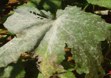
Powdery mildew is very severe on bhendi. Greyish powdery growth occurs on the under
as well as on the upper surface of the leaf causing severe reduction in fruit yield.
Pathogen
Conidia are single celled, hyaline, barrel shaped and in long chains. Cleistothecia are
globose and dark brown myceloid appendages. The asci are pedicellate, ovate or ellipsoid. The
number of ascospores is usually 2 rarely 3 per ascus. The ascospores are single celled, hyaline
and oval to sub cylindrical
Management
Spary inorganic sulphur 0.25% or Dinocap 0.1% 3 or 4 times at 15 days interval.
Vein-Clearing/Yellow Vein Mosaic : Bhendi yellow vein mosaic virus
Symptoms
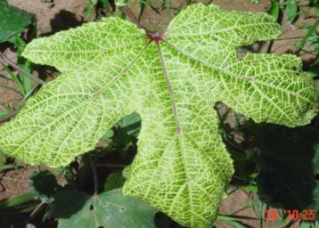
Yellowing of the entire network of veins in the leaf blade is the characteristic symptom. In
severe infections the younger leaves turn yellow, become reduced in size and the plant is highly
stunted. The veins of the leaves will be cleared by the virus and intervenal area becomes
completely yellow or white. In a field, most of the plants may be diseased and the infection may
start at any stage of plant growth. Infection restricts flowering and fruits, if formed, may be
smaller and harder. The affected plants produce fruits with yellow or white colour and they are
not fit for marketing.
Pathogen
The virus particles are 16 – 18nm in diameter.
Mode of spread
The virus is spread by whitefly.
Management
By selecting varieties resistant to yellow vein mosaic like Parbhani Kranti, Arka Abhay,
Arka Anamika, and Varsha Uphar, the incidence of the disease can be minimised. The virus is
transmitted by the whitely ( Bemisia tabaci, . Parbhani Kranti, Janardhan, Haritha, Arka Anamika
and Arka Abhay can tolerate yellow vein mosaic. For sowing during the summer season, when
the whitefly activity is high, the susceptible varieties should be avoided. Spraying
monocrotophos 1.5 ml/litre of water can restrict the disease spread. Synthetic pyrethroids should
not be used because it will aggravate the situation. It can be controlled by application of
Chlorpyriphos 2.5 ml + neem oil 2 ml lit of water.
 Phoma canker ( Phoma exigua)
Water soaked lesion appear on fruits. Black spots with irregular margin Black area
pycnidial formation. 80-90% fruit loss post harvest rot of okra pods rhizoctonia solani in brazil.
Completely rotted, the pod's typical greenish color turning brown and the infected tissues fully
covered with mycelia. Internally, immature seeds and placenta infected. Diseased tissues were
light brown to black. Externally, mycelia tend to be fluffy and lighter in color, forming a large
number of dark sclerotia on the fruit surface.
Summary Cheat Sheet
| Focus Area | Key Takeaway |
|---|---|
| Major diseases | Revise causal organisms, hallmark symptoms, and crop stage of attack. |
| Spread and survival | Link each disease with inoculum source, vector, and favorable conditions. |
| Management | Prioritize integrated management: sanitation, resistant varieties, and timely sprays. |
References
1 source • [1]
References
Class notes and standard plant pathology references
Lesson Doubts
Ask questions, get expert answers